
Figures & data
Figure 1. Archimedes stent (amg International GmbH, Winsen, Germany) has a helical structure allowing the bile flow on the outer extremity of the stent while supporting the opening of the lumen. The stent has anatomical shape, tapered tip, and proximal and distal flaps.
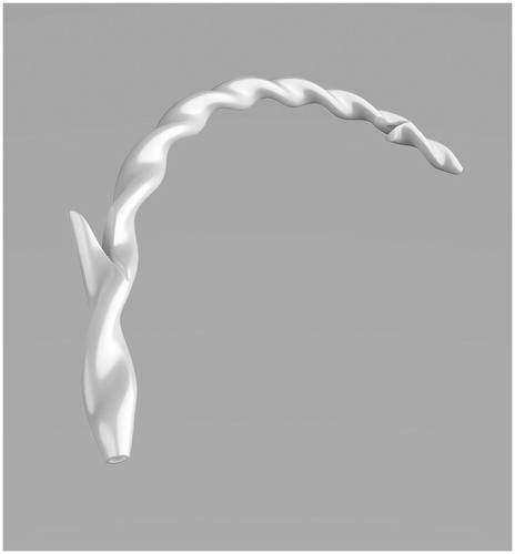
Table 1. Patient characteristics.
Table 2. Procedural characteristics.
Figure 2. The fluoroscopy views of patient (number 1) with hepaticojejunostomy anastomotic stricture (a) before and (b) after stenting with Archimedes stents.
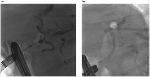
Figure 3. The fluoroscopy views of patient (number 2) with left site intrahepatic stricture (a) before and (b) after stenting with Archimedes stent.
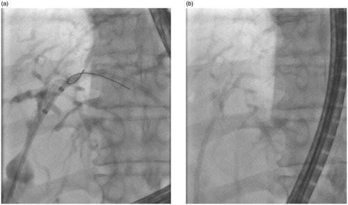
Figure 4. The fluoroscopy view of patient with hepaticojejunostomy anastomotic stricture (number 3).
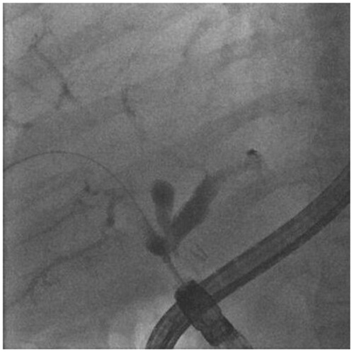
Figure 5. The fluoroscopy view of Archimedes stents of patient with hepaticojejunostomy anastomotic stricture (number 3) showing good visibility of the stents.
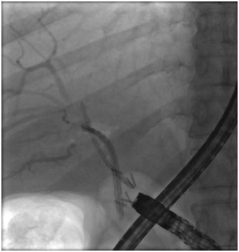
Figure 6. The endoscopy view of Archimedes stents of patient with hepaticojejunostomy anastomotic stricture (number 3).
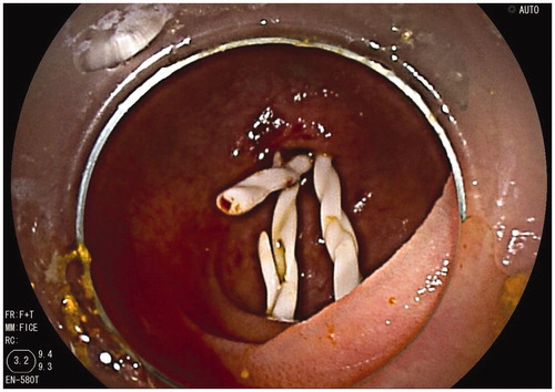
Figure 7. The fluoroscopy views of patient (number 4) with hepaticojejunostomy anastomotic stricture (a) before and (b) after stenting with Archimedes stents.
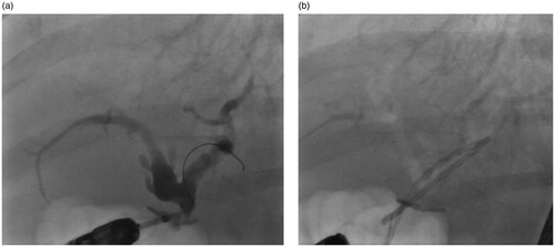
Figure 8. The fluoroscopy views of patient (number 5) with left site intrahepatic stricture (a) before and (b) after stenting with Archimedes stent.
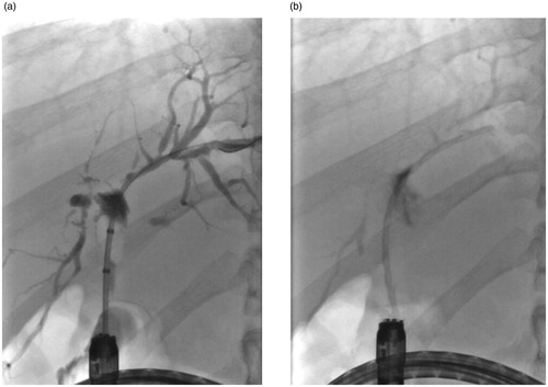
Figure 9. The fluoroscopy views of patient (number 6) with left site intrahepatic stricture (a) before and (b) after stenting with Archimedes stent.
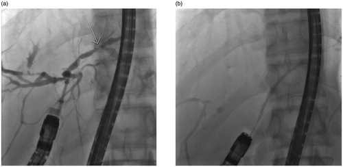
Figure 10. The fluoroscopy view of patient (number 7) with right site intrahepatic stricture before stenting.
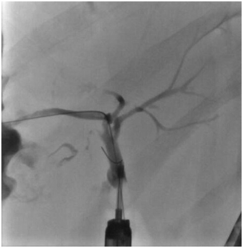
Table 3. Follow up data.