
Figures & data
Figure 1. Steatosis algorithms. Note: Steatosis algorithms (FLI, HSI, NAFLD-LFS and NAFLD Ridge score) with variables and cut off values, used for assessment of patients. Metabolic syndrome defined according to the International Diabetes Foundation criteria. NAFLD: non-alcoholic fatty liver disease; NAFLD-LFS: Non-Alcoholic Fatty Liver Disease Liver Fat Score; HSI: Hepatic Steatosis Index; FLI: Fatty Liver Index; T2DM: type 2 diabetes mellitus; BMI: Body Mass Index; ɣGT: gamma-glutamyl transferase; ALT: alanine aminotransferase; AST: aspartate aminotransferase; HDL: high-density lipoproteins; WBC: white blood cell count; HbA1c: hemoglobin A1c.
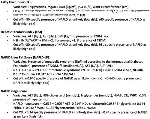
Figure 2. Advanced fibrosis algorithms. Note: Advanced fibrosis algorithms (FIB-4 and NFS) with variables and cut offs, used for assessment of patients with known steatosis or high to intermediate risk for steatosis according to steatosis algorithms. FIB-4: fibrosis-4; NFS: NAFLD Fibrosis Score; T2DM: type 2 diabetes mellitus; PC: platelet count; ALT: aspartate aminotransferase; AST: alanine aminotransferase; BMI, Body Mass Index.
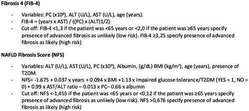
Table 1. Baseline characteristics of 350 patients with type 2 diabetes mellitus with and without probable steatosis.
Figure 3. Risk assessment by use of algorithms for high risk (light grey), intermediate risk (medium grey) and low risk (dark grey) for steatosis and advanced fibrosis in patients with type 2 diabetes mellitus. Note: Risk for steatosis and advanced fibrosis was assessed with algorithms HSI, FLI, NAFLD Ridge Score, FIB-4 and NFS. FIB-4 and NFS were calculated for all assessable patients with known steatosis or intermediate to high risk for steatosis in steatosis algorithms. NAFLD: non-alcoholic fatty liver disease; HSI: Hepatic Steatosis Index; FLI: Fatty Liver Index; FIB-4: fibrosis-4; NFS: NAFLD Fibrosis Score.
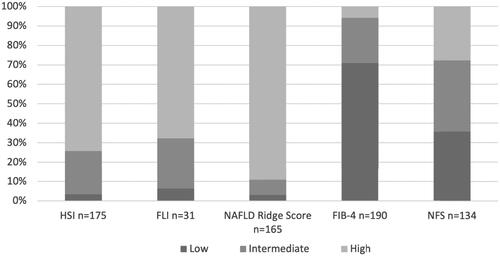
Figure 4. Panel A shows an assessment of steatosis in 350 participants with type 2 diabetes mellitus, imaging and algorithms compiled. Panel B shows an assessment of advanced fibrosis in 191 of the participants, a compilation of algorithms for advanced fibrosis. Note: Panel A: Compilation of NAFLD results for imaging and risk assessment by steatosis algorithms NAFLD-LFS, HSI, FLI and NAFLD Ridge Score for all 350 study participants. Panel B: Compilation of risk assessment with algorithms for advanced fibrosis FIB-4 and NFS for patients with known steatosis or intermediate to high risk for steatosis according to steatosis algorithm. Low risk in panels A and B is defined as low risk in all calculable algorithms, intermediate risk is defined as intermediate risk in 1–4 algorithms and none high risk assessments and high risk is defined as high risk in 1–4 algorithms. NAFLD: non-alcoholic fatty liver disease; NAFLD-LFS: Non-Alcoholic Fatty Liver Disease Liver Fat Score; HSI: Hepatic Steatosis Index; FLI: Fatty Liver Index; FIB-4: fibrosis-4; NFS: Non-Alcoholic Fatty Liver Disease Fibrosis Score.
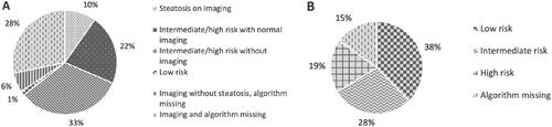
Figure 5. Results of imaging and clinical management of confirmed steatosis in this cohort of 350 patients with type 2 diabetes mellitus. Note: Present steatosis was a compilation of findings of steatosis on diagnostic imaging (US, CT and MRI) and the clinical management of known steatosis. A total of 132 (37.7%) patients had done one or more diagnostic imaging of the liver. US: ultrasound; CT: computer tomography; MRI: magnetic resonance imaging.
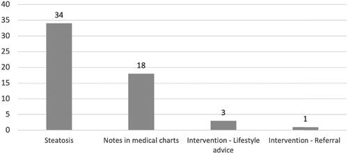
Figure 6. Screening for steatosis and advanced fibrosis in this cohort of 350 patients with type 2 diabetes mellitus according to the proposed screening algorithms from the European Association for the Study of Obesity, Diabetes, and the Liver. Note: HSI is used for assessing risk of steatosis and thereafter FIB-4 for advanced fibrosis in appliable patients. A theoretical assessment of the results of the proposed screening algorithm from the European Association for the Study of Obesity, Diabetes, and the Liver. HSI: Hepatic Steatosis Index; FIB-4: fibrosis-4.
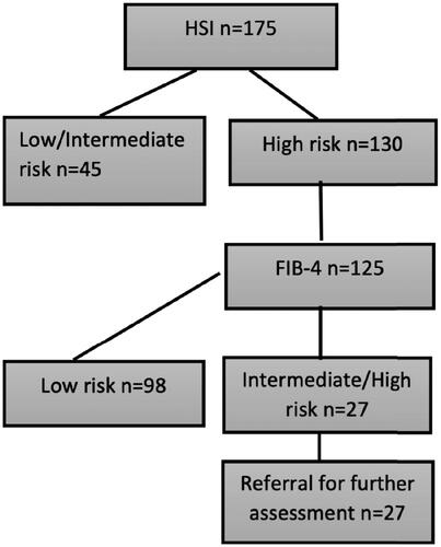
Table 2. Characteristics and comparison of groups high/intermediate risk and low risk according to algorithms (FIB-4 and NFS) for advanced fibrosis in patients with type 2 diabetes mellitus.