
Figures & data
Figure 1. Flow chart of the study profile. SEL: submucosal esophageal lesions; ESD: endoscopic submucosal excavation; STER: submucosal tunneling endoscopic resection.
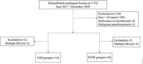
Figure 2. Endoscopic submucosal dissection for a large subepithelial esophageal lesion. (A) Endoscopic view of oval-like tumor. (B) Endoscopic ultrasonography view of the tumor. (C) Submucosal injection with fluid mixture. (D) Incision of the covering mucosa. (E) Peeling the lesions. (F) The wound after resection. (G) Closure of the mucosal incision by clips. (H) The resected specimen.
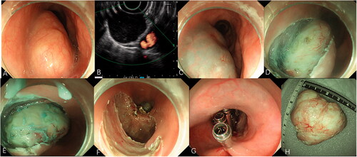
Figure 3. Submucosal tunneling endoscopic resection for a large submucosal esophageal lesion. (A) Endoscopic view of a ring-like tumor surrounding the esophagus. (B) Endoscopic ultrasonography view of the tumor. (C) A 2-cm longitudinal mucosal incision was made approximately 5 cm proximal to the SEL. (D) The submucosal tunnel is established. (E) The SEL is exposed using the submucosal tunnel technique. (F) Separating the tumor from the MP layer using the hybrid knife. (G) The mucosal entry incision is sealed with several clips. (H) Irregularly-shaped completely resected specimen (maximal diameter).
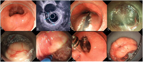
Table 1. Characteristics of 56 patients and subepithelial esophageal lesions after resection.
Table 2. Outcomes after resection in patients in submucosal tunneling endoscopic resection and endoscopic submucosal dissection.
Table 3. Comparison between the complete resection group and piecemeal resection group.
Table 4. Comparison between nonperforation group and perforation group.