
Figures & data
Figure 1. Mild atrophic gastritis, rough mucosa in the corpus, flattened rugal folds and absence of submucosal vessel.
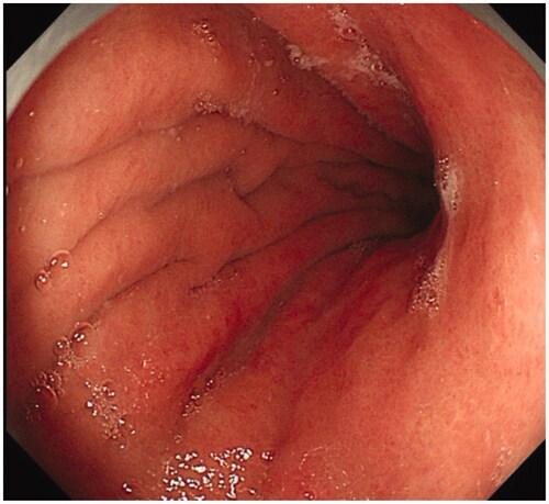
Figure 2. Moderate atrophic gastritis, thinning mucosa, flattened rugal folds and submucosal micro-vessel in the corpus.
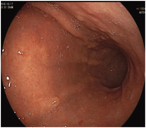
Figure 3. Severe atrophic gastritis, diminished rugal fold and submucosal grid vessel in the corpus.
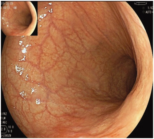
Figure 4. GHPs with well-differentiated carcinoma in the corpus were arisen from AMAG.
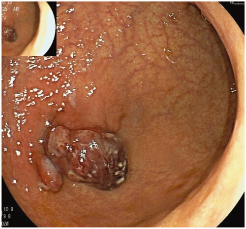
Figure 5. Multiple gastric NETs coexisted with HGD in the corpus were arisen from AMAG.
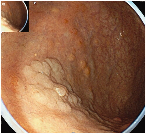
Figure 6. Two well-differentiated carcinomas in the corpus and antrum in the same patient were arisen from AMAG.
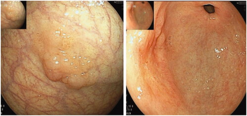
Table 1. Demographics features of AMAG patients and AMAG-associated neoplasia.
Table 2. Endoscopic characteristics of AMAG and AMAG associated gastric lesions.
Table 3. The characteristics of AMAG patients.
Table 4. Serum tests in patients of AMAG and AMAG associated neoplasia.
Table 5. Pathological characteristics of AMAG and AMAG associated gastric lesions.
Table 6. The treatments of AMAG-associated gastric lesions.