
Figures & data
Figure 1. A sagittal view of a left-sided Bochdalek hernia from a CT scan. Omental fat as well as the left kidney can be seen herniating through the posterolateral hernia defect in the diaphragm.
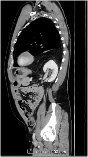
Figure 2. A sagittal view of a Morgagni hernia from a CT scan showing the defect in the anteromedial part of the diaphragm and the bowel herniated into the thoracic cavity.
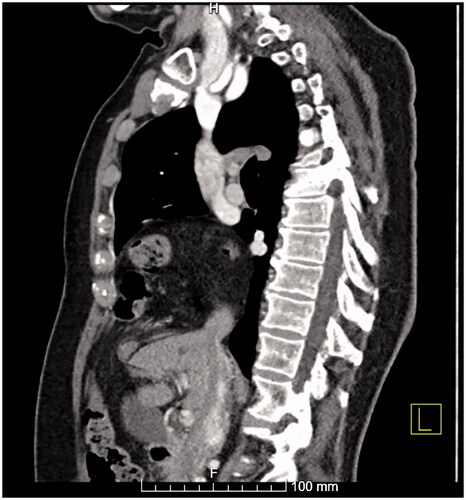
Table 1. Patient demographics.
Table 2. Hernia and operative characteristics in minimally invasive and open repairs.
Table 3. Adverse events.
Table 4. Logistic regression analysis of factors affecting complication rate.
Data availability statement
The data are available from the author. Code availability (software application or custom code).