Figures & data
Figure 1. Preoperative diagnosis of main duct IPMN. (A) MRI scan showing a universally dilated main pancreatic duct (arrows) in head, body and tail. (B) CT scan showing a 42 mm mass in the pancreatic head (arrow) and (C) an atrophic pancreatic body and tail with dilated duct (arrow).
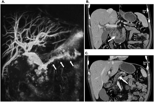
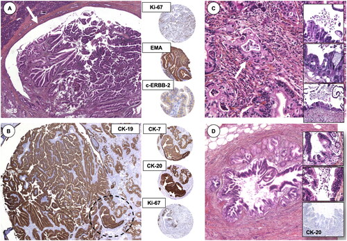
Figure 2. Histomorphological findings of an IPMN. (A) The IPMN is shown in the main pancreatic duct (arrow), with (B) positive staining for epithelial marker cytokeratin (CK)-19. Encircled area resembles the area for the immunohistochemistry markers to the right (middle column). (C) Part with invasive pancreatic adenocarcinoma and high-grade dysplasia in the pancreatic resection margin (inlets). (D) Areas of Pancreatic Intraepithelial Neoplasia (PanIN), with loss of cytokeratin-20, an epithelial marker. Presented to the annual meeting of the Norwegian Society of Pathology (Bergen, December 2004).