
Figures & data
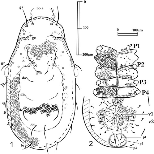
Figures 1, 2. Labidostomma motasi, male. 1, Dorsal shield. 2, Ventral view. On the epimera solely the base of setae are noted, pores are not figured.
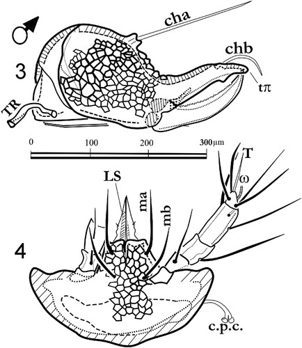
Figures 3, 4. Labidostomma motasi, male. 3, Left chelicera, internal side. 4, Infracapitulum ventral view.
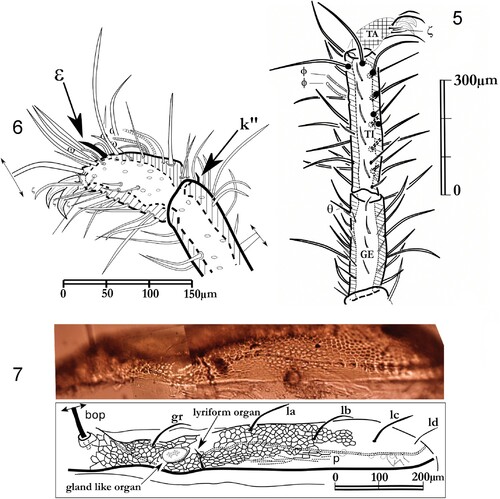
Figures 5–7. Labidostomma motasi, female. 5, Paraxial view of the tarsus I. 6, Leg I, laterodorsal view of tibia and genu. 7, Ocular zone, view of the lateral line of pores.
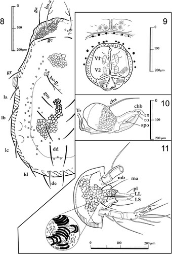
Figures 8–11. Labidostomma motasi, female. 8, Dorsal shield. 9, Ano-genital zone. 10, Right chelicera, paraxial view. 11, Infracapitulum ventral view.
Figures 12, 13. Labidostomma motasi. 12, General silhouette of the fourth leg (antiaxial view). 13, Compared lengths of the articles of the four pairs of legs.
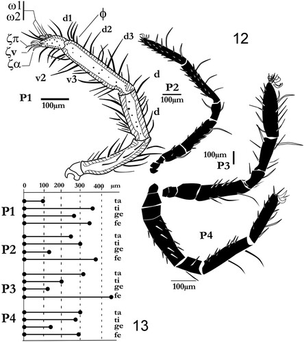
Figure 14. Labidostomma motasi, male. Lateral view of the tarsus, tibia and genu of the first of right leg showing the development of d, dorsal and v, ventral setae. Lateral line of tibial setae is noted (l’1 to l’6).
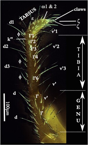
Figure 15. Diagram of the genetic distance (Cox1) of Labidostomma motasi and comparison with available data on GenBank (see text).
