
Figures & data
Figure 1 Expression levels and localization of transgenic lines as indicated by green fluorescent protein (GFP) fluorescence. (A) Schematic representation of the DNA construct, which contains the CaMV 35S RNA promoter (Pro35S), the 2805-bp region upstream of the start codon (ATG) of BOR2, the BOR2 gene sequence lacking a stop codon, GFP with a stop codon (TAA), and the nopaline synthase terminator (t-NOS). (B-D) Transgenic plants carrying the Pro35S-BOR2:BOR2-GFP or ProBOR2:BOR2-GFP constructs were grown for 5 d on solid media containing 1 µM boric acid. (B) Intensities of GFP fluorescence in roots are shown as color-coded heat maps. Upper panels show the meristem zone. Lower panels show the maturation zone. Scale bars indicate 75 µm. (C) Localization of GFP fluorescence in roots. Upper panels show the meristem zone. Lower panels show the maturation zone. (D) Localization of GFP fluorescence in epidermal cells of rosette leaves. Left upper panel shows background signal from the wild-type plants. Scale bars indicate 25 µm. (C, D).
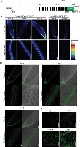
Figure 2 Vegetative growth of transgenic lines on solid media. Transgenic T3 plants were grown for 12 d on solid media containing (A) 0.03, (B) 0.1, (C) 0.3, (D) 3, and (E) 100 µM boric acid. The photographs of plants were taken on solid media (A, B, C) or on paper after transfer from solid media (D, E). Scale bars indicate 10 mm. Primary root length (F, H, J) and shoot fresh weight (G, I, K) were measured. Means ± standard deviation (SD) are shown. Student’s t-test, *p < 0.01 (n = 12–35). B, boron.
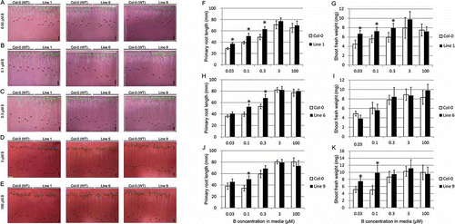
Figure 3 Reproductive growth of transgenic lines in hydroponic culture. Transgenic plants were grown for 49 d in hydroponic cultures supplemented with (A) 0.3, (B) 3, and (C) 30 µM boric acid. Upper panels in (A) show seed pods or flowers. Scale bars indicate 10 and 50 mm in the upper and lower panels, respectively.
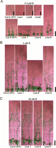
Table S1 Number of T2 transgenic line plants with hygromycin resistance. Chi-square test for 3:1 segregation of the resistant plant to the sensitive plants was done and p-values are shown.
Table S2 Segregation of T2 transgenic lines harboring Pro35S-BOR2:BOR2-GFP determined by GFP observation in T3 progenies.