Figures & data
Figure 1. An example of a high-power field of a myocyte in the left ventricle used for assessing myocyte nuclear length/size (arrows), and myocyte diameter (diamonds).
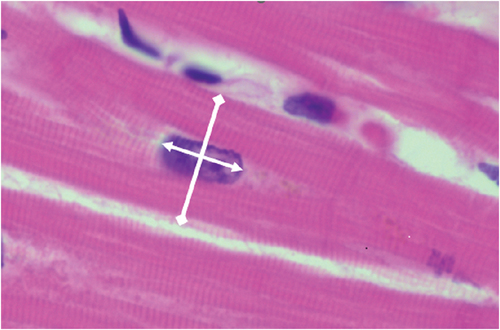
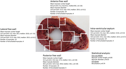
Figure 2. An illustration of statistical analysis for each case this case was a death from non-cardiac cause and the heart had cardiac hypertrophy (heart weight >600 g) without severe coronary artery atherosclerosis and other pathology. There was significant difference (ANOVA one-way p < 0.05) myocyte nuclear length only between the areas sampled. It is noted the mean myocyte diameter were above 15 µm in over 100 myocytes. The deidentified photograph is for illustration purposes and is not from the aforementioned case.