Figures & data
Figure 1. A selection of hybrid carpal arthrodesis bone plates available, demonstrating tapering of the plates at the distal end, and differences in screw-hole sizes.
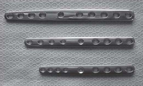
Table 1. The age, sex, breed, and weight at treatment of 14 working dogs treated using pancarpal arthrodesis, with cause of injury, and the responses of the owners to a questionnaire on the results of the surgery.
Figure 2. Dorsopalmar radiographs of a working dog with instability of the medial side of the carpus and subluxation of the antebrachioradial joint (arrow). The image on the left (L) has a stress force applied laterally, the image in the middle (N) has no stress applied, and the image on the right (M) has a stress force applied medially.
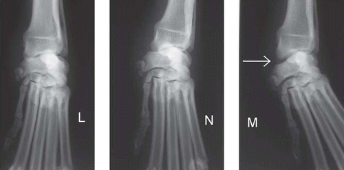
Figure 3. Post-operative mediolateral (ML) and dorsopalmar (DP) radiographs of a working dog, with application of a nine-hole 2.7/3.5-mm carpal arthrodesis bone plate.
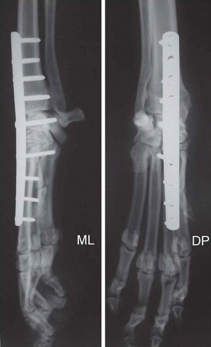
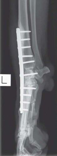
Figure 4. Mediolateral radiograph of a working dog taken approximately 6 months following surgery for pancarpal arthrodesis, demonstrating evidence of lucency around the proximal and distal ends of the carpal arthrodesis bone plate.