Figures & data
Figure 1. Trends in the prevalence of Mycobacterium bovis infection in female deer between 1993 and 2001 in central North Island areas of New Zealand. The upper series (circles) comprises all annual cohorts of adult and sub-adult females that were born before 1993 and exposed to uncontrolled M. bovis-infected possum populations. The lower series (triangles) comprises adult and sub-adult females born after the implementation of possum control measures in 1993. Adapted from Nugent (Citation2005)
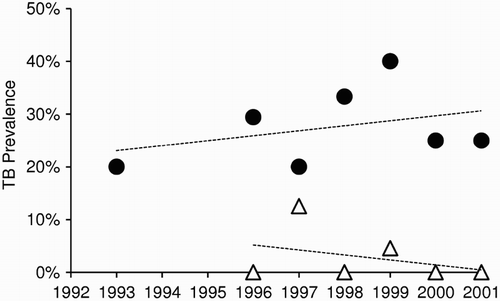
Figure 2. Mean prevalence (±SEM) of Mycobacterium bovis infection in deer killed and necropsied in the Hauhungaroa Range of the central North island of New Zealand from 1993–2003. (a) Data from the eastern side of the range where intensive possum control was applied for the first time in 1994 and (b) Data from the contiguous western side of the range where possums were not controlled during this time. Squares=0–1 year-olds, triangles=1–2 year-olds, circles=>2 year olds. Adapted from Nugent (Citation2005)
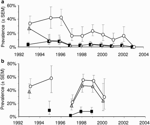
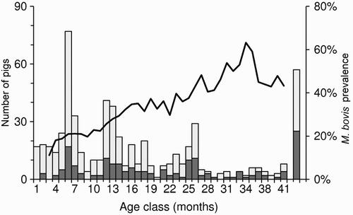
Figure 3. Five-month running average prevalence of Mycobacterium bovis infection (line) and sample size (columns) for 592 pigs aged 1–41 months that were necropsied between 1994 and 2008 in three regions of New Zealand. The far right bar represents all adult pigs >3.5 years old (∼10% of the sample). The dark shading represents the number of tuberculosis-positive animals. Adapted from data presented in Nugent and Whitford (Citation2008b), with permission