Figures & data
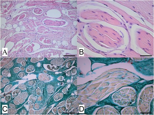
Figure 1. Images from surgery undertaken to remove a spherical, deep dermal mass, about 3 cm in diameter, from the upper neck of a 17-year-old castrated male red deer (Cervus elaphus): A) the site of the mass on the left side of the deer’s neck at the angle of the jaw; B) the spherical mass after removal.
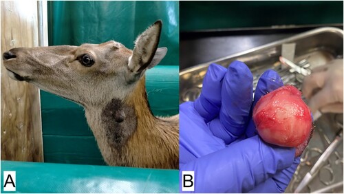
Figure 2. Photomicrographs of a dermal mass removed from the neck of a red deer (Cervus elaphus) showing A) a representative area of the mass located within the deep dermis and comprising well differentiated muscle bundles surrounded and often widely separated by an extensive collagen stroma (H&E; bar = 75 µm) and B) detail of several muscle bundles within the mass and, in the upper right of the image, an area of collagen stroma (H&E; bar = 35 µm). Slides shown in C) (low magnification; bar = 75 µm) and D (high magnification; bar = 35 µm) are stained with Masson’s trichrome stain, and show the different components of the mass, with muscle bundles staining pale orange-red while the surrounding collagen stroma stains blue-green.