Figures & data
Figure 1. (A) Photograph of lesions on the mammary gland of an 18-year-old Thoroughbred mare, showing visible enlargement of the left gland with multiple exudative ulcerated lesions and discharge from the left teat. (B) Ultrasound images of the left (top) and right (bottom) mammary glands showing areas of mixed echogenicity with microlobulated margins and an abrupt interface between the mass and normal mammary tissue (circle).
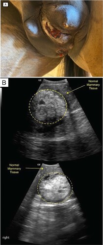
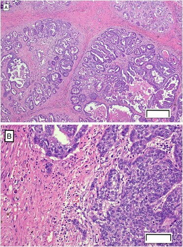
Figure 2. Photomicrographs of sections of mammary tissue from a Thoroughbred mare, showing (A) epithelial cells forming tubules and papillary projections within the neoplasia of the mammary tissue (H&E; bar = 200 µm) and (B) clusters of cells invading the basement membrane (H&E; bar = 50 µm).