Figures & data
Table 1. Signalment, diagnostic findings, details of case management and outcome in four dogs diagnosed with protothecosis in New Zealand.
Figure 1. Photographs of a dog (Case 4) with (a) multiple foot pad ulcerations, (b) an ulcerated swelling of the left elbow and (c, d) firm nodular swellings within the skin of the metacarpi with ulceration of the swelling of the right metacarpus (c).
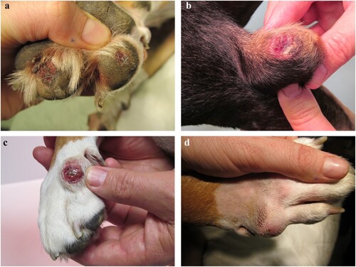
Figure 2. Photomicrograph of section from the (a) colon of Case 1 and (b) skin of Case 4 showing endosporulating algal organism (arrows) with internal septation in both locations and empty theca (arrow heads) in the section of skin (H&E; bar = 30 μm). These are magnified in the insets.
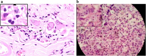
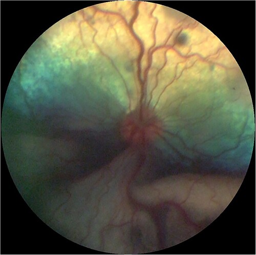
Figure 3. Fundic photograph of a dog with ocular protothecosis showing multifocal hypo-reflective areas of active chorioretinitis and an opaque, ventral, exudative retinal detachment. Image courtesy of Marnie Ford, Animal Eye Care, Melbourne, Australia.