Figures & data
Figure 1. Ultrasonographic images of the coelomic cavity of a female West Coast green gecko (Naultinus tuberculatus) showing a) sagittal view of a single follicle (F) in the coelom on admission; b) transverse view of two follicles (F) in the coelom on admission; c) sagittal view of two follicles (F) in the coelom after 6 months of hospitalisation; d) oblique view of four follicles (F) in the coelom after 6 months of hospitalisation, suggestive of pre-ovulatory follicular stasis.
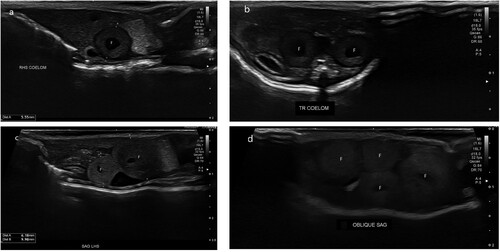
Figure 2. Intra-operative images of the exploratory coeliotomy of a female West Coast green gecko (Naultinus tuberculatus) with preovulatory follicular stasis, showing a) the ovary and two follicles exterior to the paramedian incision; b) the technique used for expressing the yolk prior to folliculectomy. The gecko is under a transparent surgical drape.
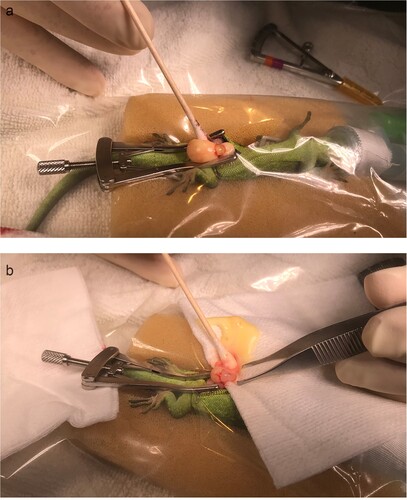
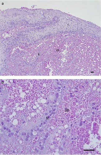
Figure 3. Histology of the resected ovarian follicle, showing: a) relatively thick theca (t), and the single cuboidal-columnar layer of follicular cells (fc) are displaced, possibly as a result of the surgery but this may be secondary to POFS; b) the follicular cells (fc) and mature yolk protein (yp) with very few vesicles present and no evidence of inflammation or degeneration (H&E, scale bars = 50 µm).