

Abstract
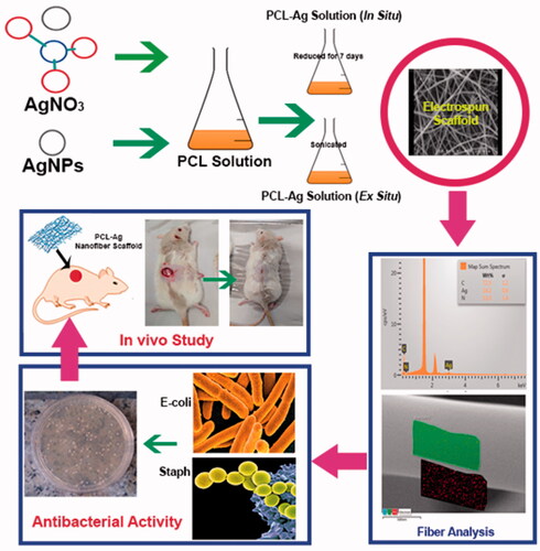
Poly(ɛ-caprolactone)-silver nanocomposite were prepared by two main methods of metal-polymer nanocomposite preparation approaches, in situ and ex situ methods, by electrospinning. Effective parameters on electrospun nanofibers were optimized by Design Expert, CCD method. Using in situ and ex situ methods, effect of incorporating silver nanoparticle into Poly(ɛ-caprolactone) on conductivity, average fiber diameter, morphology of nanofiber mats and antibacterial properties were investigated. According to Field Emission Scanning Electron Microscopy (FESEM) and Energy-Dispersive X-ray Spectroscopy (EDX) analysis, average fibers diameters decreased by increasing incorporated Ag nanoparticles. Conductivity results confirmed the higher content of silver nanoparticles in in situ samples. Antibacterial test was performed against two bacteria types: Staphylococcus-aureus and Escherichia-coli. Due to higher content and smaller size of Ag nanoparticles incorporated in nanofibers, silver-loaded PCL electrospun nanofibers obtained by in situ method showed higher antibacterial efficiency in comparison to the ex situ samples. Moreover, Ag particles had more effects on Staphylococcus aureus due to sensitive membrane of this type of bacteria. In vivo study has proved that in situ samples improved the wounds on Wistar rat lateral part of body skin. Antibacterial and in vivo results showed that PCL-Ag nanofiber mats obtained by in situ method are suitable for medical applications.
Graphical Abstract
To achieve low average fibers diameters, in situ method was more applicable than ex situ one.
Antibacterial properties of fibers mat prepared by in situ method was more effective than ex situ one against both Staphylococcus aureus and Escherichia coli bacteria.
Effectiveness of AgNPs in both in situ and ex situ methods on Staphylococcus aureus was more than Escherichia coli.
In situ and ex situ nanofibers have proved to be more effective in in vivo test and showed better performance than pure PCL nanofiber scaffolds.
HIGHLIGHTS
Acknowledgments
The authors thank the Iran Nanotechnology Initiative Council (INIC) for the financial support of this work Linkage project (46160).