Figures & data
Figure 1. Matching (a) textbook (From Marieb EN: Human Anatomy & Physiology, San Francisco, 2001, Benjamin Cummings, used by permission of Pearson Education, Inc.) and (b) cadaver roll-over images of the location of the heart in the mediastinum of the thorax showing the aorta. A comparison of (a) and (b) shows that the ‘empty spaces’ that are frequently included between structures for clarity in textbook drawings do not actually exist in the body and that structures are often less perfect in shape when seen in situ.
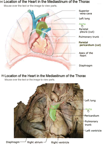
Figure 2. Cadaver image dissected to reveal the major abdominal arteries. (a) Roll-over highlight of abdominal aorta. (b) Roll-over highlight with the abdominal aorta still emphasized but now adding in two branches, the common iliac arteries.
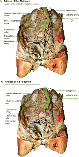
Figure 3. Corresponding (a) textbook (From Marieb EN: Human Anatomy & Physiology, San Francisco, 2001, Benjamin Cummings, used by permission of Pearson Education, Inc.) and (b) cadaver roll-over images of the right upper limb and thorax highlighted to allow the arterial pathway of blood flow to the wrist to be followed.
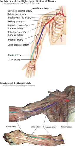
Figure 4. Rotating roll-over image of the skull with the sphenoid bone highlighted so that it can be followed as the skull is manipulated to be viewed at (a) 0°, (b) 45°, and (c) 90° of rotation. (d) The grouping function has been activated to allow all of the facial bones to be viewed simultaneously at 45° of rotation.
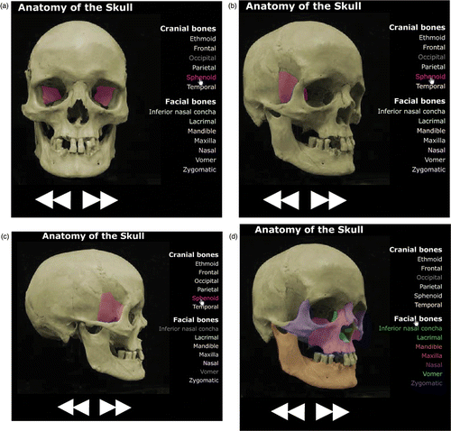
Figure 5. Fade-through textbook (From Marieb EN: Human Anatomy & Physiology, San Francisco, 2001, Benjamin Cummings, used by permission of Pearson Education, Inc.) image of the heart with a sliding bar allowing the transparency of the more superficial of the image components to be adjusted between 0 and 100%.
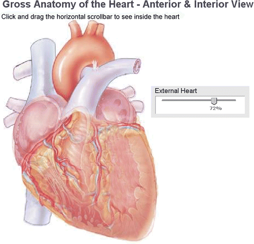
Figure 6. Reusable learning applets. (a) Partially completed drag-and-drop image of the heart. (b) Self-testing image of the upper respiratory tract in which the epiglottis has been highlighted and its corresponding label correctly entered. (reprinted by permission of Pearson Education, Inc.).
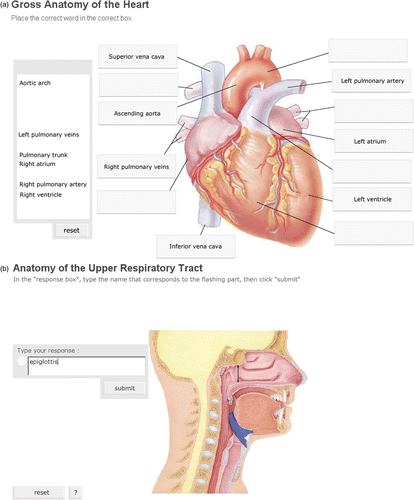
Table 1. Comparison of class distribution of final grades for students who had access to online learning tools (LT Group) or did not have access to these tools (No-LT Group) throughout the academic term.*