Figures & data
Table 1. Summary of patient population (n = 70) and clinical characteristics
Figure 1. Immunohistochemistry assay for Napsin A expression. (a) Negative expression in healthy oral mucosa. (b) Napsin A has strong, diffuse granular cytoplasmic expression in pulmonary adenocarcinoma. (DAB) Scale bar = 400 µm
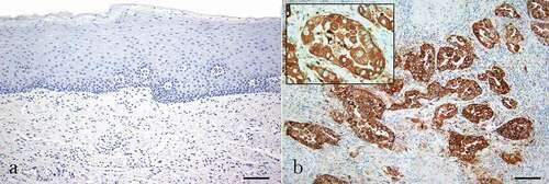
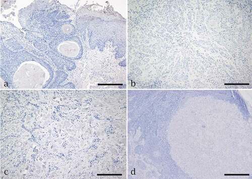
Figure 2. Immunohistochemistry assay for Napsin A expression in oral squamous cell carcinoma (OSCC). Negative expression of napsin A is seen in (a) a well-differentiated tumor and edge of the epithelium, (b) a moderately differentiated tumor, (c) a poorly differentiated tumor and (d) OSCC metastasis to neck lymph node. Scale bar = 200 µm for a, b,c. Scale bar = 50 µm for d