Figures & data
Figure 1. Flow diagram of the process used to select eligible studies.
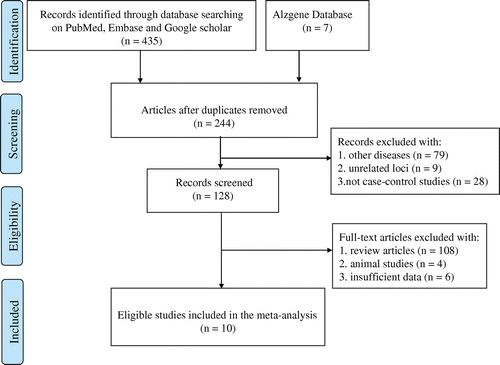
Table 1. The baseline characteristics of all the included studies in this meta-analysis.
Table 2. Quality assessment scheme for included studies (Newcastle–Ottawa Scale).
Figure 2. Forest plots of MTHFR A1298C polymorphism and the risk of Alzheimer’s disease in five genetic models: A: the allelic model (C vs. A), B: the homozygous model (CC vs. AA), C: the heterozygous model (CA vs. AA), D: the dominant model (CC + CA vs. AA), E: the recessive model (CC vs. CA + AA), and F: the allelic model (C vs. A) in cumulative meta-analysis by publication year.
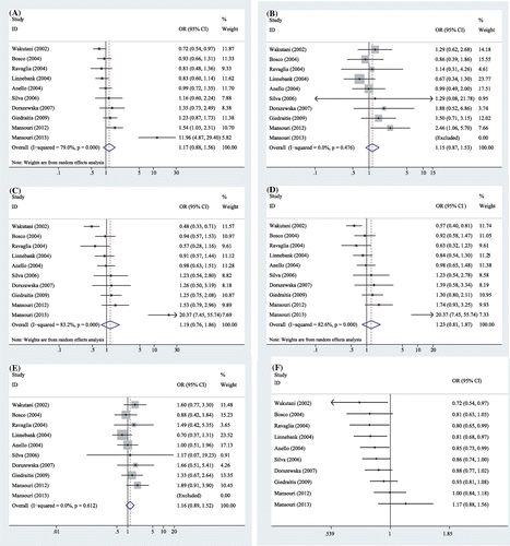
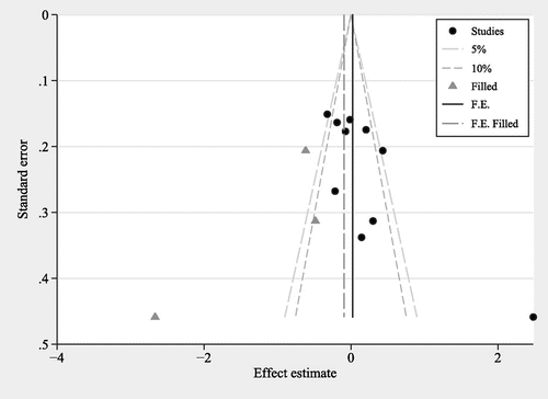
Figure 3. Funnel plot for the allelic model (C vs. A) to analyze publication bias between MTHFR A1298C polymorphism and AD risk.
Notes: Studies: included studies in this meta-analysis, 5 and 10%: contour lines aiding the interpretation of the funnel plot, Filled: filled estimated studies using the trim and fill method, F.E.: the combined effect with estimated studies not filled. and F.E.Filled: the combined effect with estimated studies filled.