
Figures & data
Table 1. Ingredients of control Diet A and presumed high-risk Diet B, both based on AIN-76A.
Figure 1. An example of the very few discrete (or well defined) aberrant crypt foci (ACF) observed in animals on Diet B in Study 3. Note the slit-like lumens likely indicative of a dysplastic histology. (The mucosal field is 1.7 × 0.8 mm.)
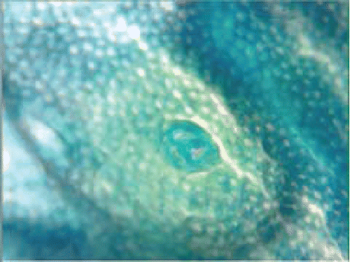
Figure 2. Two examples of heterogeneous (or ill-defined) aberrant crypt focus (ACF) as observed on the colonic mucosal surface after Bouin's fixation in Study 2. The ACF are the foci of large, darkly staining enlarged crypts bordering an infolding pattern over what appears to be a lymphoid follicle. Faint outlines of the openings of small crypts are seen at the center of both ACF. (The mucosal fields are 1.7 × 0.8 mm and 2.7 × 1.3 mm.)
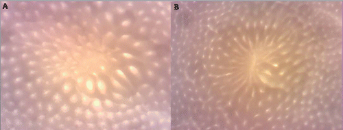
Figure 3. Two examples of heterogeneous (or ill-defined) aberrant crypt focus (ACF) as observed on the colonic mucosal surface following formalin fixation and staining with Methylene Blue in Study 3. Again, a faint outline of the openings of small crypts can be seen at the center of the ACF. We suggest that these changes follow the loss of clusters of crypts and are an early response to the loss of adjacent crypts (The mucosal fields are 1.7 × 0.8 mm.).
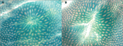
Figure 4. An example of the histological appearance at low and high power of an inflammatory/hyperproliferative focus observed in Study 3. On histologic examination of the colons that had been “Swiss rolled,” the foci were noted by their narrow caliber, mucin, and Goblet cell-depleted crypts (yellow arrow in ), and then by the frequent mitotic figures (red arrows in ), the damaged luminal surface of the aberrant crypt focus, and the many acute and chronic inflammatory cells. The large crypts, so evident on the topologic en face preparation, were not as evident in this histological section.
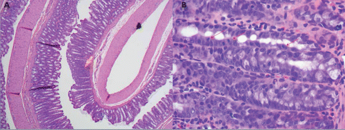
Table 2. Metabolic, biochemical and clinical measures for rats after 14 weeks on Diet A and B (Study 1).
Table 3. Colonic mucosal surface measures for rats after 40 weeks on Diet A and B (Study 2).
Table 4. Metabolic, biochemical, clinical, and colonic mucosal surface measures for rats after 14 weeks on Diet A and B with decreased fluorescent light (UV) (Study 3).