Figures & data
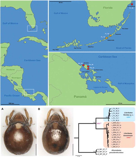
Table 1. GenBank accession numbers for COI and 18S rRNA gene sequences comprising all specimens included in genetic investigations. Sequences generated in this study appear in bold.
Figure 1. Maps illustrating the records of new species in the western Caribbean. Left vertical map provides an overview of the area with white frames indicating the position of the regions given in the two horizontal maps. Red circles represent records of Thalassozetes balboa sp. nov. and orange circles identify occurrence of Litoribates floridae sp. nov.; codes in white refer to the sampling location given in the Materials and Method section.
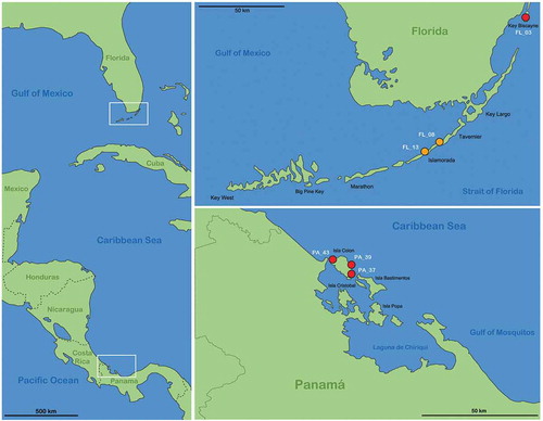
Figure 2. Graphic illustration of measured continuous variables shown on simplified drawings of Thalassozetes barbara. (a) Dorsal aspect: bl – body length, dPtI – distance between pedotecta I, db – distance between bothridia, ll – lenticulus length, dnr – distance notogastral ridges, nwda – notogastral width on level of seta da, nwdm – notogastral width on level of seta dm, nwdp – notogastral width on level of seta dp. (b) Ventral aspect: cl – camerostome length, cw – camerostome width, dre1 – distance between ridges on epimeron 1, efl – epimeral fovea length, efw1 – epimeral fovea width anterior part, efw2 – epimeral fovea width posterior part, dcg – distance between camerostome and genital orifice, dac3 – distance between acetabula 3, gl – genital orifice length, gw – genital orifice width, al – anal opening length, aw – anal opening width.
Figure 3. Litoribates floridae sp. nov. adult (a) dorsal view; (b) ventral view, distal leg segments omitted; (c) lateral view.
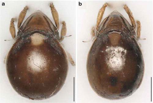
Figure 4. Photographs (stacked stereomicroscopic images) of Litoribates floridae sp. nov. adult (a) dorsal view; (b) ventral view; (c) lateral view. Scale bars 100 µm.
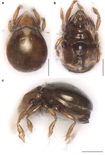
Figure 5. Litoribates floridae sp. nov. adult mouthparts (a) right rutellum, ventral view; (b) left pedipalp, paraxial view; (c) left chelicera, antiaxial view.
Table 2. Leg chaetome and solenidia for the new species.
Figure 6. Litoribates floridae sp. nov. adult left legs, antiaxial view (a) leg I; (b) leg II; (c) leg III; (d) leg IV.

Figure 7. Thalassozetes balboa sp. nov. adult (a) dorsal view; (b) ventral view, distal leg segments omitted; (c) lateral view.
Figure 8. Photographs (stacked stereomicroscopic images) of Thalassozetes balboa sp. nov. adult (a) dorsal view; (b) ventral view; (c) lateral view. Scale bars 100 µm.
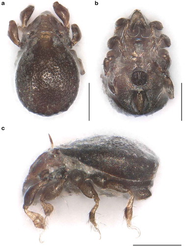
Figure 9. Thalassozetes balboa sp. nov. adult mouthparts (a) right rutellum, ventral view; (b) left pedipalp, paraxial view; (c) left chelicera, antiaxial view.
Figure 10. Thalassozetes balboa sp. nov. adult left legs, antiaxial view (a) leg I; (b) leg II; (c) leg III; (d) leg IV.
Table 3. Univariate statistics for Caribbean Litoribates species.
Table 4. Loadings of the two principal components PC 1 and PC 2 gained from PCA on data of L. bonairensis and L. floridae sp. nov.
Figure 11. Scatter plots of PCA of L. bonairensis from Bonaire and L. floridae sp. nov. from Florida. Open symbols refer to females; filled symbols refer to male specimens. For better display of sexual dimorphism, each gender is given in a separate hull.
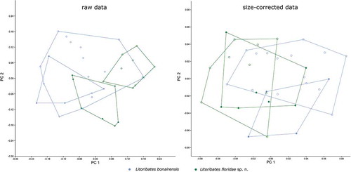
Table 5. Univariate statistics for Caribbean Thalassozetes species.
Table 6. Loadings of the two principal components PC 1 and PC 2 gained from PCA on data of T. barbara and T. balboa sp. nov.
Figure 12. Scatter plots of PCA of T. barbara from Barbados and T. balboa sp. nov. from three different locations in Panama. Codes refer to sample sites given in the Materials and Method section; open symbols refer to females; filled symbols refer to male specimens.
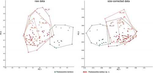
Figure 13. Bayesian inference tree of three fortuyniid species based on COI sequences constructed by means of MrBayes applying the GTR+I+G model. Posterior probabilities for main nodes are shown above branches. Sequences obtained from GenBank are marked by *. For details, see . ** Mean of intra- (blue, red) and interspecific (violet) uncorrected p-distances given in per cent.
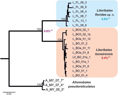
Figure 14. Bayesian inference tree of fortuyniid and selenoribatid taxa based on 18S rRNA sequences (1811 bp) constructed by means of MrBayes applying the GTR+I+G model. Posterior probabilities (>80) are shown near nodes. Sequences obtained from GenBank are marked by *. For details, see .
Figure 15. Photographs (stacked stereomicroscopic images) of female adult Litoribates specimens used for morphometric investigations. (a) Litoribates floridae sp. nov., Florida; (b) Litoribates bonairensis, Bonaire. Scale bars 100 µm.