Figures & data
Figure 1. a. Intraoperative image of a tiger undergoing SILS™ port ovariectomy. The Ligasure Atlas™ and 10 mm inner cannula are to the left, the 5 mm telescope and camera are in the middle cannula, and the 5 mm Endo Grasp™ is to the right. b. Image of the SILS™ port with one 10 mm and two 5 mm inner cannulas.
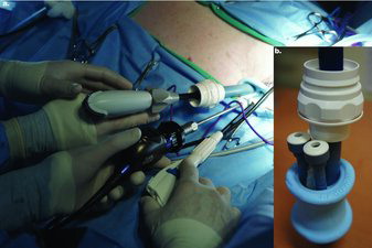
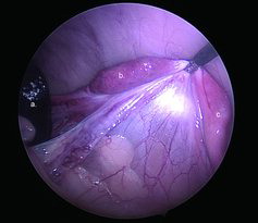
Figure 2. Intraoperative image of the ovarian pedicle in one of the tigers. a. Spleen. b. Ovary. c. Uterine horn. Notice the open ovarian bursa, which was observed with both ovaries of both tigers.