1,368
Views
5
CrossRef citations to date
0
Altmetric
Review Articles

The aryl hydrocarbon receptor and food allergy

, &
Pages 94-107 | Received 06 May 2013, Accepted 07 May 2013, Published online: 08 Jun 2013

Figures & data

Figure 1. Schematic overview of mechanisms involved in AhR activation. Scheme 1: After ligation of the AhR with an agonist, the ligand:AhR complex translocates to the nucleus where it dimerizes with ARNT. Then, hsp90, hsp23 and the X-associated protein 2 dissociate from the complex and the ligand:AhR:ARNT complex binds to DREs, resulting in the transcription of genes. Scheme 2: Proteolytic degradation of the AhR by the 25S proteosome. Schemes 3 and 4: AhR-mediated induction of the AhRR reduces the formation of the ligand:AhR:ARNT complex. Scheme 5: AhR ligands are metabolically depleted by CYP450 enzymes. Adapted from Mitchell and Elferink (Citation2009).
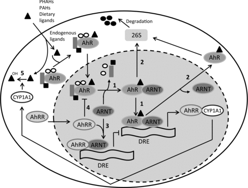
Figure 1. Schematic overview of mechanisms involved in AhR activation. Scheme 1: After ligation of the AhR with an agonist, the ligand:AhR complex translocates to the nucleus where it dimerizes with ARNT. Then, hsp90, hsp23 and the X-associated protein 2 dissociate from the complex and the ligand:AhR:ARNT complex binds to DREs, resulting in the transcription of genes. Scheme 2: Proteolytic degradation of the AhR by the 25S proteosome. Schemes 3 and 4: AhR-mediated induction of the AhRR reduces the formation of the ligand:AhR:ARNT complex. Scheme 5: AhR ligands are metabolically depleted by CYP450 enzymes. Adapted from Mitchell and Elferink (Citation2009).

Figure 2. Structures of synthetic, endogenous and dietary AhR ligands.

Figure 2. Structures of synthetic, endogenous and dietary AhR ligands.

Figure 3. Differentiation of CD4+ cells into different effector T cells; T helper 1 (Th1), T helper 2 (Th2), T helper 17 (Th17) or regulatory T (Treg) cells. Differentiation of these T cell subsets is mediated by the interaction of dendritic cells with naïve CD4+ T cells, the presence of specific cytokines and mediators and cell-type specific transcription factors. By secreting specific cytokines, effector T cells mediate their regulatory or activating properties as shown on the right. Adapted from Jetten (Citation2009).
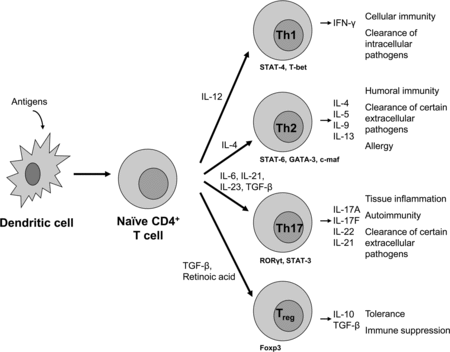
Figure 3. Differentiation of CD4+ cells into different effector T cells; T helper 1 (Th1), T helper 2 (Th2), T helper 17 (Th17) or regulatory T (Treg) cells. Differentiation of these T cell subsets is mediated by the interaction of dendritic cells with naïve CD4+ T cells, the presence of specific cytokines and mediators and cell-type specific transcription factors. By secreting specific cytokines, effector T cells mediate their regulatory or activating properties as shown on the right. Adapted from Jetten (Citation2009).

Table 2. Effects of AhR activation on Th17 and Treg cells in various disease models as described in text.

Figure 4. Schematic representation of an IgE-mediated allergic reaction to food allergens. In normal individuals, presentation of processed proteins and peptides by DCs to naïve T cells will lead to the induction of high-dose tolerance (induced by T cell clonal anergy and apoptosis) or low-dose tolerance (active suppression of the immune reaction via induction of Treg cells). However, in certain individuals, presentation of a food protein by DCs on MHCII may result in the induction and activation of Th2 cells. By secreting cytokines (e.g. IL-4, IL-5, IL-13), these Th2 cells stimulate B cells to produce allergen-specific IgE which is distributed systemically. This allergen-specific IgE binds to high affinity IgE receptors (FcϵRI) present primarily on mast cells and basophils. This process is known as allergic sensitization. Re-exposure (challenge) to the food antigen results in crosslinking of the IgE bound on FcϵRI, which provokes degranulation of mast cells and basophils and the release of mediators (such as histamines, cytokines, leukotrines, prostaglandins and platelet-activating factor). This leads to a variety of clinical effects.
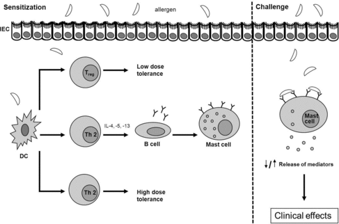
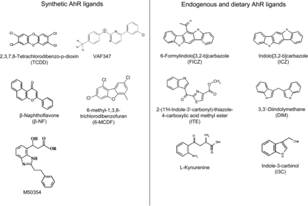
Figure 4. Schematic representation of an IgE-mediated allergic reaction to food allergens. In normal individuals, presentation of processed proteins and peptides by DCs to naïve T cells will lead to the induction of high-dose tolerance (induced by T cell clonal anergy and apoptosis) or low-dose tolerance (active suppression of the immune reaction via induction of Treg cells). However, in certain individuals, presentation of a food protein by DCs on MHCII may result in the induction and activation of Th2 cells. By secreting cytokines (e.g. IL-4, IL-5, IL-13), these Th2 cells stimulate B cells to produce allergen-specific IgE which is distributed systemically. This allergen-specific IgE binds to high affinity IgE receptors (FcϵRI) present primarily on mast cells and basophils. This process is known as allergic sensitization. Re-exposure (challenge) to the food antigen results in crosslinking of the IgE bound on FcϵRI, which provokes degranulation of mast cells and basophils and the release of mediators (such as histamines, cytokines, leukotrines, prostaglandins and platelet-activating factor). This leads to a variety of clinical effects.

Reprints and Corporate Permissions

Please note: Selecting permissions does not provide access to the full text of the article, please see our help page How do I view content?

To request a reprint or corporate permissions for this article, please click on the relevant link below:

Academic Permissions

Please note: Selecting permissions does not provide access to the full text of the article, please see our help page How do I view content?

Obtain permissions instantly via Rightslink by clicking on the button below:

If you are unable to obtain permissions via Rightslink, please complete and submit this Permissions form. For more information, please visit our Permissions help page.