Figures & data
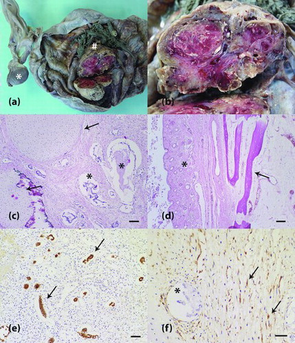
Figure 1. Ovarian teratoma in an equine fetus. (a) The right ovary shows a solid mass (hash) within a cystic structure. The left ovary is observed and exhibits a blackened appearance (asterisk). (b) Magnification from (a). A solid mass reveals a whitish to reddish lobulated cut surface. (c) Neoplastic tissue contains well-differentiated tubular arrangements of neoplastic epithelial cells (asterisk) and cartilaginous matrix (arrow). H&E staining is shown, and the scale bar represents 100 μm. (d) Other components of the neoplasm were pavimentous stratified keratinizing epithelium, pilous follicles (asterisk) and osseous matrix (arrow). H&E staining is shown at 100x magnification. (e) Tubular arrangements of epithelial cells with cytoplasmic immunopositivity for cytokeratin (arrow). Peroxidase system (Advance HRP Enzyme, DakoCytomation, Carpinteria, CA, USA). The cells were stained with the anti-cytokeratin AE1AE3 primary antibody and counterstained with Harris’ hematoxylin. The scale bar represents 50 μm. (f) Vimentin-positive neoplastic mesenchymal cells (arrow). Negativity for vimentin can be observed in neoplastic epithelial cells (asterisk). Peroxidase system (Advance HRP Enzyme, DakoCytomation, Carpinteria, CA, USA). The cells were stained with an anti-vimentin primary antibody and counterstained with Harris' hematoxylin. The scale bar represents 30 μm.