Figures & data
Table 1 Age, sex and breed of examined dogs (nos. 1–6 group of young dogs and nos. 7–36 group of elderly dogs).
Figure 1 Congo red staining (semi-quantitative evaluation), magnification 100x: (A) grade + (low number of the positive blood vessels of Congo red positive vessels in the total number of vessels in field), (B) grade ++ (moderate number of Congo red positive vessels in the total number of vessels in field).
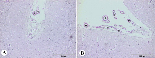
Figure 2 Congo red staining of the brain of aged dogs: (A) amyloid in the wall of meningeal blood vessel (arrow), frontal section; (B) amyloid in the wall of meningeal blood vessel (arrow), frontal section, under the polarized light; (C) amyloid in the wall of meningeal blood vessels of cerebellum (arrow), under polarized light.
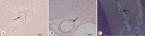
Table 2 Semi-quantitive evaluation of the cerebral amyloid angiopathy in different tissue sections (Congo red staining).
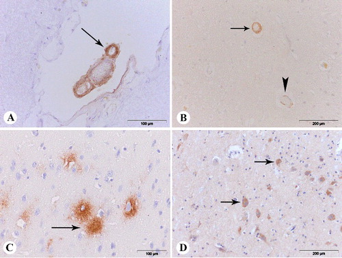
Figure 3 (A) β-amyloid deposits in the wall of meningeal blood vessels (arrow), anti-Aβ1-42, frontal section; (B) β-amyloid deposits in the wall of parenchymal blood vessels (arrow – severe type of the Vonsattel scale and score 4 of the Mountjoy scale, arrowhead – mild type of the Vonsattel scale and score 1 of the Mountjoy scale), anti-Aβ 1-14, frontal section; (C) diffuse plaque (arrow), anti-Aβ 1-42 parietal section; (D) intracellular β-amyloid deposits (arrow), anti-Aβ 1-14, parietal section.