Figures & data
Table 1. Hematological data of an injured gopher tortoise with systemic inflammation. Reference values are minimum and maximum values from 13 healthy gopher tortoises (Taylor & Jacobson Citation1982). For reference values, absolute numbers of leukocytes were calculated based on percentage of each leukocyte type times the mean total white blood cell (WBC) count. NR = not reported.
Figure 1. Heterophil projections in the blood film of an injured gopher tortoise at day of admission. The projections are in the same color as heterophil granules and visible as rows of individual or fused heterophil granules (up to 30 m in length) that were arranged in semi-circular or circular formations. ×100 objective. Wright-Giemsa stain.
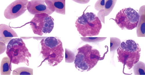
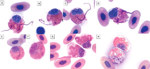
Figure 2. Heterophil projections in blood films of an injured gopher tortoise during 2.5 months of treatment (A-E; G). The projections are visible in mature (round nucleus and low nuclear to cytoplasmic ratio) and in immature heterophils (oval or elongated nucleus, larger nucleus compared to mature stage, immature granules, larger cell size). Mild toxicity is visible as mild degranulation, cytoplasmic basophilia, and/or vacuolation. A-B, E: mature heterophils, mild degranulation, and slight cytoplasmic basophilia; C-D, F-G: immature heterophils, mild degranulation, and slight cytoplasmic basophilia; H upper cell: mature heterophil; H bottom cell: immature heterophil, mild degranulation, cytoplasmic basophilia, and vacuolation. ×100 objective. Wright-Giemsa stain.