
Figures & data
Table 1. Highlights of this rabies virus review.
Table 2. Journey of rabies virus.
Figure 1. Gross and histopathological lesions in rabies. (A) Marked congestion of blood vessels in sulci of swollen cerebral hemispheres. (B) Massive perivascular infiltration with mononuclear cells (lymphocytes and macrophages) around dilated blood vessels in white matter of camel brain. H&E x400. (C) Dense eosinophilic and sharply outlined Negri bodies (arrow) of various sizes in the cytoplasm of intact neuron in camel brainstem section. H&E x400. (D) Brain section showing degenerated neurons, severe cuffing with mononuclear cells and edema. A Negri body is also visible in the degenerated neuron (arrow) having mild diffuse gliosis. H&E x200.
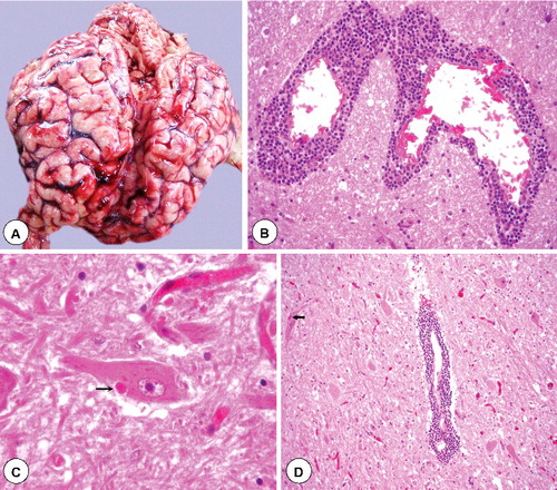
Figure 2. Diagnostic techniques for rabies. (A) Brain impression smear showing oval or round magenta coloured Negri bodies (arrow) in the cytoplasm of neuron. Seller's stain x1000. (B) Intense brown colour signals of rabies virus antigen in the cytoplasm of pyramidal neurons in experimentally infected mice brain with challenge virus standard (CVS) strain of rabies virus. IHC-DAB-MH x200. (C) Brain impression smear showing bright apple green dusty fluorescent signals of rabies virus antigen in the cytoplasm of neurons in a spotted deer. Inset showing plenty of specific signals in the cytoplasmic processes and stroma of neuron. dFAT x200. (D). Bright apple green fluorescent signals of rabies virus antigen in the smear of saliva in a dog. dFAT x200.
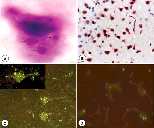
Table 3. Type of contact, exposure and recommended post-exposure prophylaxis.