
Figures & data
Figure 1. Structure of Nipah virus.
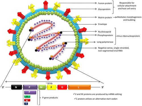
Figure 2. Transmission of the Nipah virus. 1. Fruit bats acts as natural reservoir of Nipah viruses. Fruit bats with NiV feeds on date palm sap. Virus can survive in solutions that are rich in sugar, viz., fruit pulp. 2. Virus transmitted to human through the consumption of date palm sap. 3. Fruit bats of Pteropus spp. which are NiV reservoirs visited such fruit trees and got opportunity to naturally spill the drop containing virus in the farm to contaminate the farm soil and fruits. 4. Contaminated fruits are consumed by pigs and other animals. Pigs act as intermediate as well as amplifying host. Combination of close surroundings of fruiting trees, fruits-like date palm, fruit bats, pigs and human altogether form the basis of emergence and spread of new deadly zoonotic virus infection like Nipah. 5. Pork meat infected with NiV are exported to other parts. 6. Consumption of infected pork can act as a source of infection to human. 7. Close contact with NiV affected human can lead to spread of NiV to other persons.
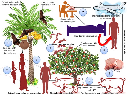
Figure 3. Phylogenetic analyses of sequences of Nipah Virus (NiV) strains from different countries (Bangladesh, Cambodia, India, Malaysia, and Thailand). (A) Phyloanalysis based on complete G gene (1809 bp) and (B) Phyloanalysis based on complete N gene (1599 bp). Tree created with maximum likelihood method with 1,000 bootstrap replicates. Scale bars indicate number of sequence changes corresponding to illustrated branch length. Major two NiV clades are mentioned in the side bar as BD (Bangladesh) and MY (Malaysia).
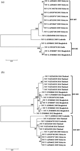
Figure 4. Pathogenesis of NiV. 1. NiV can be seen in the epithelial cells of the bronchiole in the initial stage of infection. 2. NiV antigen can be detected in bronchi and alveoli. 3. Inflammatory mediators are activated as a result of infection to the airway epithelium. 4. Virus is disseminated to the endothelial cells of the lungs in the later stage of the disease. 5, 6. Virus enter the blood stream followed by dissemination, either freely or in host leukocyte bound form, reach brain, spleen and kidneys. 7. Two pathways are involved in the process of viral entry into the central nervous system (CNS), via hematogenous route and anterogradely via olfactory nerve nerves. 8. The blood brain barrier (BBB) is disrupted and IL-1β along with tumor necrosis factor (TNF)-α are expressed due to infection of the CNS by the virus which ultimately leads to development of neurological signs. Red font shows the symptoms in human.
Figure 5. Vaccine platforms for NiV. 1. Recombinant measles virus (rMV) vaccine that expresses envelope glycoprotein of NiV has been found to be effective vaccine candidate. 2. A recombinant vaccine based on vesicular stomatitis virus (replication-competent) has been developed in recent years encoding a glycoprotein of NiV. 3. Nipah virus-like particles (NiV-VLPs) composed of three NiV proteins G, F and M derived from mammalian cells have been produced and validated as vaccine in BALB/c mice. 4. Immunoinformatic advances have been utilized for developing peptide-based NiV vaccine by prediction and modeling of T-cell epitopes of NiV antigenic proteins.
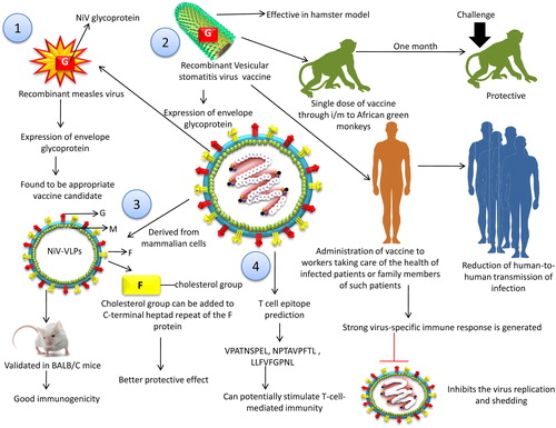
Table 1. Different vaccine strategies available for Nipah virus (NiV).