Figures & data
Figure 1. Horizontal ultrasonographic scan of the right eye. The eye shows a “hourglass” shape with a larger echoic anterior lesion (arrows) that continues in a posterior rounded mixed-echoic portion, attributable to the vitreous chamber and the fundus (empty arrow heads). Legend: A = anterior; P = posterior; L = lateral; M = medial.
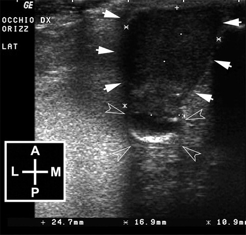
Figure 2. A: Histopathological section of the tumor mass stained with hematoxylin and eosin (magnification: 4×): Iris is infiltrated by a densely cellular neoplastic proliferation (asterisk). B: Histopathological section of the tumor mass stained with hematoxylin and eosin (magnification 40×): The neoplastic cells appear pleomorphic, rarely pigmented (arrowheads), with high nucleus-to-cytoplasm ratio and show numerous mitoses (arrows).
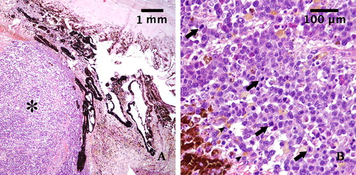
Figure 3. A: Neoplastic cells expressing Melan-A (3,3′-Diaminobenzidine indirect immunohistochemistry. Hematoxylin in counterstain) with strong label intensity in some cells. (original magnification 40×). B: Neoplastic cells expressing S100 (3,3′-Diaminobenzidine indirect immunohistochemistry. Hematoxylin in counterstain) with strong label intensity diffuse in cellular proliferation. (original magnification 40×).
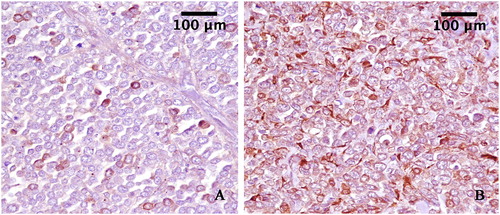
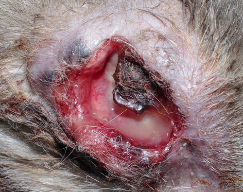
Figure 4. The cat, 14 months after surgery. Neoplastic tissue invading orbital space and developing over the site of the exenteration scar, associated with cutaneous necrosis and ulceration, can be observed.