Figures & data
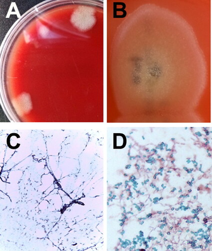
Figure 1. Changes in C-reactive protein (CRP), ratio of urine protein to creatinine (UPC), and body temperature during the therapeutic monitoring period after the diagnosis of infective endocarditis (IE) in a 8-year-old, spayed, female Maltese dog. After the diagnosis of IE, oral administration of the antibiotics showed no improvement in CRP (reference range, 0.1–1 mg/L), UPC (reference range, 2–5 mg/L), body temperature, and clinical signs. Intravenous administration of the antibiotics resulted in blood culture negative, and there were improvements in body temperature, UPC, and CRP levels.
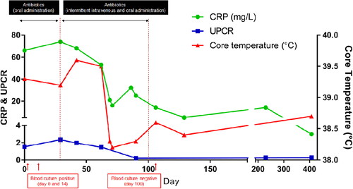
Figure 2. Two-dimensional echocardiographic images of the right parasternal long-axis four-chamber view (A), right parasternal long-axis five-chamber view (B), right parasternal short-axis view (C), thickened anterior leaflet of mitral valves (arrow), and the noncoronary cusp of aortic valves (arrow head) with irregular and hyperechoic vegetative lesions in a 8-year-old, spayed, female Maltese dog. Left atrial thrombus was confirmed (asterisk). Echocardiogram images of the right parasternal long-axis four-chamber view (D) and right parasternal long-axis five-chamber view (E) after 4 months follow-up. The thickening of anterior leaflet of mitral valves (arrow) was still identified, but vegetative lesions of the noncoronary cusp of aortic valve (arrow head) were not found. Decreased size of thrombus was confirmed in the left atrium. LV, left ventricle; RV, right ventricle, LA, left atrium, RA, right atrium; Ao, aorta.
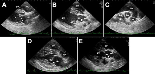
Figure 3. Identification of bacterial isolates from an 8-year-old, spayed, female Maltese dog with infective endocarditis. Initial bacterial colony morphology (A) on the blood agar plate after enrichment in blood culture bottles under aerobic and anaerobic incubation conditions. Close-up appearance of characteristic Bacillus amyloliquefaciens colonies (B) grown on blood agar plates (see white, fluffy, wool-like appearance of the colony). Gram-staining (C) and spore-staining (D) of the bacteria grown on the nutrient agar surface (C) and in nutrient broth (D). The results of the bacterial strain showed that the bacteria were somewhat entangled, forming filamentous branches (C). Numerous malachite green-stained Bacillus spores were formed in the broth (D).