Figures & data
Table 1. Details of the outbreaks in wild boars.
Figure 1. Gross alterations in ASFV infected tissues. (a) Enlarged and congested lymph nodes. (b) Extensive petechial haemorrhages in the subcapsular area of the kidney.
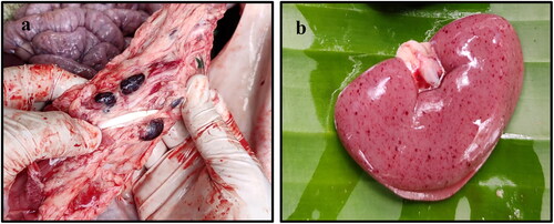
Figure 2. Histopathological examination of ASFV infected tissues. (a) Intestine showing infiltration of lymphocytes in the submucosa (magnification 40 times, (b) Liver showing necrosis (yellow arrow) with lymphoplasmacytic infiltration (black arrow)(40×), (c) Kidney showing aggregation of mononuclear cells (yellow arrow) with haemorrhages (40×), (d) Lung showing accumulation of inter and intra alveolar aggregation of mononuclear cells, predominantly macrophages (40×), (e) Lymph node showing depletion of lymphocytes from lymphoid follicle (yellow arrow) with accumulation of necrotic debris (10×), and (f) Spleen showing depletion of lymphocytes from the splenic corpuscles (red circle)(10x).
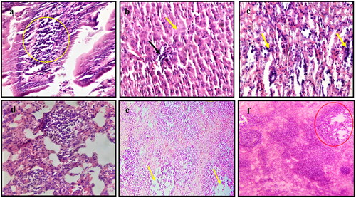
Figure 3. Phylogenetic tree of ASFV based on partial B646L gene. The ASFV strains detected in wild boars and domestic pigs (highlighted with red circle) formed clade with genotype-II strains (dark blue colour clade) of ASFV.
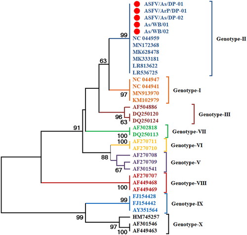
Figure 4. Timeline of ASFV outbreaks. Timeline of suspected ASFV outbreaks in Assam and Arunachal Pradesh which might be responsible for the probable transmission of ASFV from domestic pigs to wild boars of Assam.
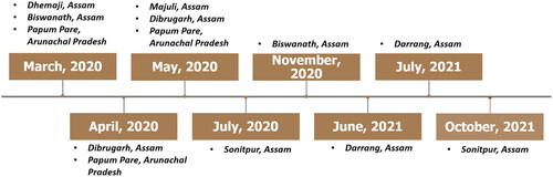
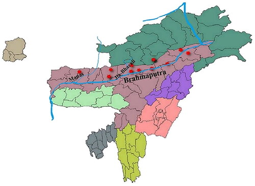
Figure 5. Map of North-eastern states of India representing ASFV outbreaks in the national parks (Manas and Nameri) and the places near the river Brahmaputra where outbreaks were reported. The tributaries (Manas and Jia-Bharali rivers) of Brahmaputra that passes through the two national parks are also depicted in the map. Outbreaks at different locations are highlighted with red marker.