
Figures & data
Table 1. Correlation between CaChPV-1 detection and age of affected dogs.
Figure 1. Phylogenetic topology of Carnivore chaphamaparvovirus-1 (CaChPV-1) based on full-length nucleotide sequences, non-structural gene 1 (NS1), and viral capsid gene 1 (VP1). The CaChPV-1 obtained from dogs presented a monophyletic clade (blue dot box) separating it from feline chaphamaparvovirus. The CaChPV-1 sequences obtained in this study (red triangles) were separately grouped into two lineages that were clustered with the CaChPV-1 sequences found in China and Canada, supported by phylogenetic analysis of the NS1 and VP1 genes. Bars indicate nucleotide substitution per site.
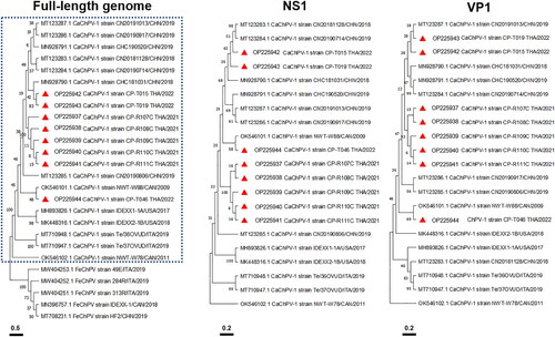
Figure 2. Carnivore chaphamaparvovirus-1 infection. Puppy no. 2. Duodenum (A&B). (A) Focally extensive lymphoplasmacytic duodenitis (arrows) and villous necrosis. Lymphocyte and plasma cell infiltration in intestinal submucosa (inset). Hematoxylin & eosin (H&E) staining. (B) The lumen of several blood vessels within the remnant villous structures are filled with granular to fibrillar materials (arrows), highlighted by deep blue color (inset). Phosphotungstic acid-hematoxylin (PTAH) staining. Puppy no. 3. Lung (C&B). (C) Interstitial pneumonia and diffuse pulmonary congestion. The alveolar lumen of small bronchiolar airways is filled with pools of inflammatory cells, and lining type II pneumocytes segmentally exhibit tombstoning hypertrophy. (D) These inflammatory cells are predominantly composed of lymphocytes, neutrophils, and foamy macrophages. H&E. Bars indicate 100 µm (A&B), 200 µm (C), and 50 µm (D).
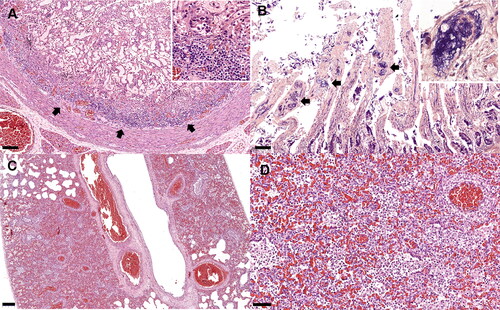
Figure 3. Carnivore chaphamaparvovirus-1 infection. In situ hybridization. Puppy no. 2. Intestine (A-C). Mesenteric lymph node (D). Lung (E&F). (A) Large amounts of carnivore chaphamarparvovirus-1 (CaChPV-1) nucleic acid in the duodenum. (B, C) The CaChPV-1 DNA is present in the superficial mucosa of intestinal villi. The CaChPV-1 nucleic acid is primarily detected within the nuclei of supporting mesenchymal cells and endothelial cells of villous capillaries. (D) CaChPV-1 DNA is detected in single lymphocytes and histiocytes in the mesenteric lymph node. (E) Large amounts of CaChPV-1 nucleic acid are multifocally present in pulmonary parenchyma and small blood vessels (inset). (F) CaChPV-1 hybridization signals are primarily present in the nuclei of infiltrated histiocytes and lymphocytes in alveoli and present in lining endothelial cells of alveolar capillaries (arrow). Bars indicate 500 µm (A), 100 µm (B&E), 20 µm (C&F), and 10 µm (D).
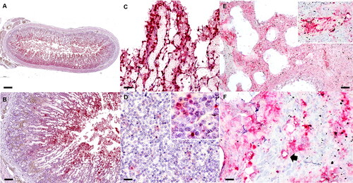
Table 2. Detection of CaChPV-1 in various organs of necropsied canine puppies with fatal enteric disease.
Supplemental Material
Download MS Word (16.9 KB)Data availability statement
All the data supporting our findings is contained within the manuscript. Eight full-length coding sequences of the CaChPV-1 have been deposited in NCBI GenBank under accession numbers OP225937–OP225944.