
Figures & data
Figure 1. The methods of gross pathological and histological morphometrical analysis. (A) The methodology of the gross measurements of the studied hearts that were conducted on the transverse section of the ventricular wall at the level of the upper third of the ventricle (below the valvular ring). (B) The methodology of the histological morphometric measurements of intramyocardial coronary arteries as proposed by Falk et al. (Citation2006); picro sirius red stain, 200× magnification. AA: arterial area; aid: measured arterial internal diameter; AID: calculated arterial internal diameter; awt: measured arterial wall thickness; AWT: calculated arterial wall thickness; DWTR: arterial diameter-to-wall thickness ratio; HS: heart surface; IVS: interventricular septum; LAR: arterial lumen-to-area ratio; LVID: left ventricular internal diameter; LVLS: left ventricular lumen surface; LVS: left ventricular surface; LVW: left ventricular wall; LVWS: left ventricular wall surface; RVID: right ventricular internal diameter; RVLS: right ventricular lumen surface; RVW: right ventricular wall.
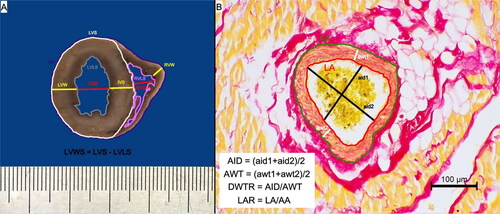
Table 1. The method of semi-quantitative histological evaluation of cardiomyocyte degeneration, myocardial inflammatory infiltration and myocardial disarray in the ventricular specimens of the examined animals based on Janus et al. (Citation2016) and Biasato et al. (Citation2015).
Table 2. The age, sex, body weight and breed of the studied animals.
Table 3. The results of the pathomorphological examination of the studied hearts.
Figure 2. The cross section of the ventricular walls in the control (A), FHT (B) and HCM (C) groups. The patient’s heart weight and their respective body weight: 15 g and 4.3 kg (A), 23 g and 5 kg (B), 39 g and 4 kg (C).
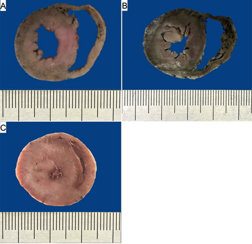
Figure 3. The results of the pathomorphological examination of the studied hearts. The results are presented as mean ± 95% confidence intervals. Values presenting significant differences are marked with asterisk (* or **). IVS: interventricular septum diameter; LAA: left atrial appendage; LV: left ventricle; LVW: left ventricular wall diameter; LVWs: left ventricular walls; RVW: right ventricular wall diameter.
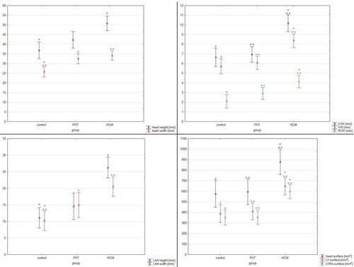
Table 4. The results of the semi-quantitative histopathological analysis of the studied hearts.
Table 5. The results of quantitative histopathological examination in the studied groups.
Figure 4. The histological image of the intramural arteries in the examined cats. Picro sirius red stain; 200× magnification. (A, D, G) Control group; (B, E, H) FHT group; (C, F, I) HCM group; (A–C) left ventricular free wall; (D–F) interventricular septum; (G–I) right ventricular free wall. IVS: interventricular septum; LVW: left ventricular free wall; RVW: right ventricular free wall.
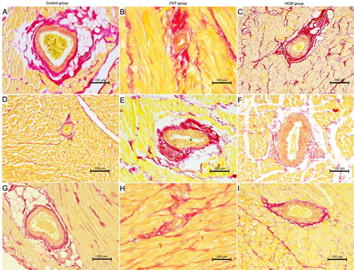
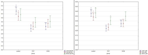
Figure 5. The results of the measurements of coronary arteries in the studied hearts. The results are presented as mean ± 95% confidence intervals. Values presenting significant differences are marked with asterisk (* or **). DWTR: arterial diameter-to-wall thickness ratio; IVS: interventricular septum; LAR: arterial lumen-to-area ratio; LVW: left ventricular wall; RVW: right ventricular wall.
Supplemental Material
Download MS Word (18.8 KB)Data availability statement
The research data are available at the main author after reasonable request