
Figures & data
Figure 1. (A) Binding profile and (B) cPD-1/cPD-L1 neutralizing profile of four mouse anti-cPD-L1 mAbs. Data are presented as mean ± SD.
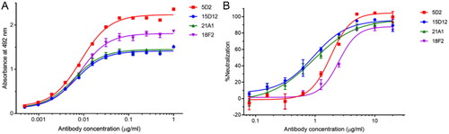
Table 1. Summary characteristics of four purified mouse anti-cPD-L1 mAbs.
Figure 2. SPR sensorgrams of purified mouse anti-cPD-L1 mAb clone (A) 5D2, (B) 15D12, (C) 18F2, and (D) 21A1.
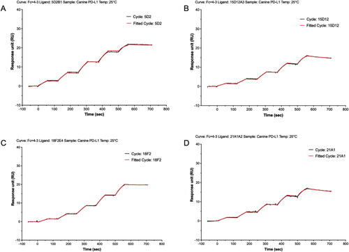
Table 2. Binding kinetics data of mouse anti-cPD-L1 mAbs against its target.
Figure 3. Canine PD-L1 immunohistochemical staining in cutaneous T-cell lymphoma. (A) Clone 5D2. (B) Clone 15D12. (C) Clone 18F2. (D) Clone 21A1. 400×. IHC. Bar = 10 µm.
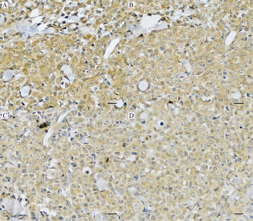
Figure 4. Immunohistochemistry staining of cPD-L1-positive tissues of clone 5D2. (A) 100% of neoplastic cells in squamous cell carcinoma, (B) oral malignant melanoma, (C) soft tissue sarcoma, and (D) tubular mammary carcinoma presents strong membranous and cytoplasmic staining for cPD-L1 (400×). IHC. Bar = 10 µm.
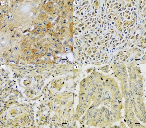
Table 3. Immunohistochemical results of mouse anti-cPD-L1 mAbs in five cases of cutaneous T-cell lymphoma, tubular mammary carcinoma, and soft tissue sarcoma.