
Figures & data
Figure 1. Gallbladder wall thickness in dogs with hypoalbuminemia. The serum albumin level of the dogs with normal gallbladder wall thickness (<2 mm; Group A) was shown in blue dots and with gallbladder wall thickening (Group B and C) was shown in orange dots. The serum albumin level of the dogs in Group A (1.82 ± 0.33 g/dl) and the dogs in Groups B and C (2.10 ± 0.17 g/dl) was not significantly different (p = 0.14). No correlation was present between the serum albumin level and the gallbladder wall thickness (p = 0.12).
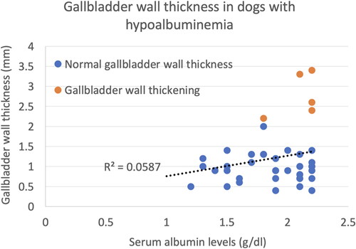
Figure 2. Ultrasound images of gallbladder wall thickening in dogs with hypoalbuminemia. (A) A mild and diffuse homogeneously hyperechoic gallbladder wall thickening is present. This dog was diagnosed with lymphoma. (B) A mild and diffuse gallbladder wall thickening with a 3-layer appearance. There are thin inner and outer hyperechoic layers and a central hypoechoic layer. This dog was diagnosed with hepatic metastatic carcinoma. (C) A mild and diffuse gallbladder wall thickening with a 3-layer appearance. Hyperechoic lacy striations are present within the central hypoechoic layer. This dog was diagnosed with the caudal vena cava obstruction due to neoplasia associated thrombosis with hepatic congestion and ascites.
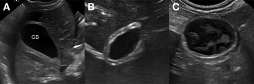
Table 1. Ultrasonographic characterics and other information of the dogs with gallbladder wall thickening (>2 mm) and hypoalbuminemia.
Data Availability Statement
The data that support the findings of this study are available from the corresponding author, MM, upon reasonable request.