Figures & data
Figure 1. The FMDV infected and recovered calves showing higher serum levels (p < 0.0001) of glucose (a), cTn-I (b), T3 (c), T4 (d), and reduced levels of TSH (e) as compared to control calves. The datas were analyzed by one-way ANOVA with bonferroni post hoc test using GraphPad prism 8.0.1 statistical software.
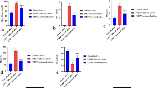
Figure 2. Gross and histopathological changes in various tissues of calves naturally infected with FMDV. a) Heart, cattle calf, 2.5 M. The ventricular myocardium showing necrotic grey to white streaks of variable size and shape giving ‘tigroid heart’ appearance. b) cross-sectional view of heart showing necrotizing myocarditis. c) Thymus, cattle calf, 4 M: swollen thymus with a few pin-head size blood spots on its surface. d) Lymph node, buffalo calf, 4 M. Cut surface of the lymph node showing severe congestion. e) Tongue, cattle calf, 3 M. Ballooning degeneration of stratum spinosum accompanied with severe inflammatory reaction. Hematoxylin and eosin (HE), X100. f) Heart, buffalo calf, 4 M. Heart showing acute interstitial necrotizing myocarditis characterized by marked infiltration of mononuclear cells (arrow). HE, X200. g) Lymph node, calf, 4 M. Depleted lymphoid follicles (arrow) in the cortex. HE, X100. h) Thymus, cattle calf, 4 M. Severe engorgement of blood vessel with the infiltration of inflammatory cells in the capsule of thymus. HE, X100. h) Small intestine, cattle calf 5 M. Enteritis showing marked infiltration of inflammatory cells in the lamina propria. HE, X100. j) Spleen, cattle calf, 4 M. Depleted lymphoid follicles in the white pulp region. HE, X100. k) Pancreas, buffalo calf, 4 M. Multifocal interstitial pancreatitis characterized by infiltration of inflammatory cells (arrow) surrounding the pancreatic acinar cells. HE, X100. l) Thyroid, cattle calf, 4 M. Presence of few inflammatory cells with scant colloid in the thyroid follicle. HE, X100.
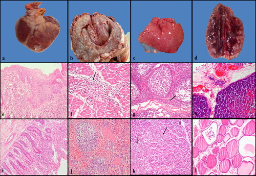
Table 1. Frequency of FMDV induced lesions in different organs of 37 young cattle and bubaline calves affected with FMDV under natural condition.
Figure 3. The immunolocalization of FMDV antigen in various tissues of FMDV affected calves showing a) tongue showing distinct intense cytoplasmic immunostaining in the stratum basal and spinosum layer of cattle calf. The density of reaction is much higher in the stratum basal layer, IHC, X100. b) Distribution of FMDV antigen diffusely in multifocal manner in the necrosed cardiac muscle fibers of buffalo calf, IHC, X100. c) The macrophages infiltrating the tonsillar crypt epithelium showing the presence of FMDV antigen, IHC, X100. Note the presence of viral antigen in the necrotic cellular debris. d) Thymus showing cytoplasmic immunoreactivity for FMDV antigen in hassall’s corpuscles of cattle calf, IHC, X200. e) Pancreas showing high amount of FMDV antigen in the islets of Langerhans of buffalo calf. Note the moderate cytoplasmic immunoreactivity in the few pancreatic acinar cells, IHC, X100. f) Thyroid showing intense immunostaining for FMDV antigen in the follicular cells of cattle calf, IHC, X100. g) Adrenal gland showing diffuse granular strong cytoplasmic staining in zona reticularis layer of the adrenal cortex of cattle calf, X200. h) The small intestinal epithelial cells as well as the intraepithelial inflammatory cells showed the presence of FMDV antigen of buffalo calf, IHC, X100. i) The tubular epithelial cells of the kidney in cattle calf showing cytoplasmic immunopositivity for FMDV antigen, IHC, X100.
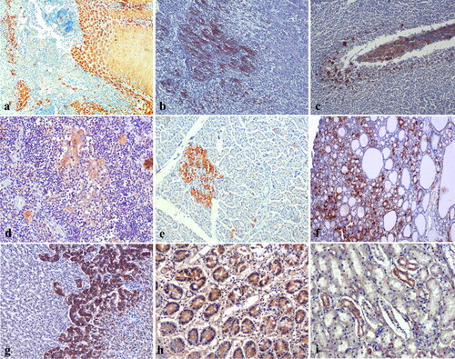
Table 2. Severity and localization of the immunohistochemical findings in various tissues of cattle and buffalo calves naturally affected with FMDV.
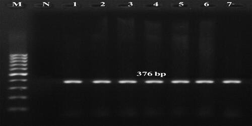
Figure 4. Molecular detection of FMDV type A by multiplex RT-PCR in tissue samples of calves. Lane M–100 bp + DNA ladder, N-negative control, Lane 1–7 amplicons positive for Type A (376 bp), L1-Tongue, cattle calf, case 1, L2-Heart, cattle calf, case 4, L3- Tongue, buffalo calf, case 8, L4- Heart, case 9, L5- Tongue, buffalo calf, case 9, L6- Heart, buffalo calf, case 9, L7-Heart, cattle calf, case 7.