
Figures & data
Figure 1. Histopathological features of DCH infection in lymphoma tissue of cats. (A, E-J) Sections from case no. 3. (A) The lymphoid follicles are diffusely expanded by large neoplastic lymphocytes separating coarse fibrovascular stroma. Neoplastic lymphocytes have moderate amounts of cytoplasm with distinct cellular borders. The nuclei are 2 times larger than red blood cells, round, and finely stippled, containing a prominent nucleolus (square box is indicated for the inset). (B-D) DCH qPCR-positive lymphoma section of case no.6. (B) Diffuse, generalized cytoplasmic IHC staining for CD79. (C) Prominent nuclear IHC staining for pax-5 in multiple neoplastic cells. (D) Cytoplasmic IHC staining for CD3 in some single cells in lymphoma tissue. (E) In situ hybridization (ISH) for DCH DNA staining (red precipitates) in lymphoma tissue revealed intensely intranuclear staining in diffuse round cells (square box is indicated for the inset). (F) Prominent nuclear staining for DCH hybridization in large, pleomorphic round cells. No DCH labeling was observed within the small round cells. (G) Dual DCH ISH (red precipitates) and CD79 IHC (green color) revealed marked, diffuse staining in the nucleus and cytoplasm of neoplastic cells, respectively. (H) Higher magnification reveals intense DCH labeling (red precipitates) in the nucleus of the cells that were cytoplasmically stained with CD79 IHC (green color). (I) Negative control using unrelated probe. No hybridization signals were present in the DCH qPCR-positive lymphoma section that was incubated with the FBoV-3 ISH probe. (J) Negative control using DNase-treated section. No hybridization was present in the DCH qPCR-positive lymphoma section treated with DNase prior to incubation with the DCH probe. (K) Negative control for dual ISH/IHC labeling. Cytoplasmic IHC CD79 staining of the DCH qPCR-positive lymphoma section incubated with the FBoV-3 probe. Bars indicate 180 µm for A, B, D and E; 80 µm for F; 120 µm for C, G, I, and J; and 20 µm for H and K.
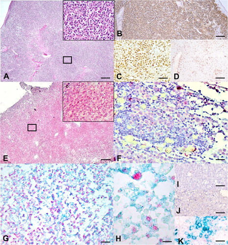
Table 1. Detection of DCH DNA in blood and lymph node tissues of investigated cats.
Table 2. Morphological characteristics of lymphoma cats and DCH DNA detection in paraffin-embedded lymphoma tissues.
Table 3. Histological characteristics of DCH-positive feline lymphoma cases.
Data availability statement
All data generated or analysed during this study are included in this published article.