
Figures & data
Figure 1. Ex vivo intestinal mucosal abrasion model. The intestine is mounted on the black platform (1) with wheels underneath. It is pulled forward by a pulley winding system (2). The intestinal segment is cautiously stretched in 2 directions with a gripping mechanism (3), the bi-axonal tension is measured with force meters (4). The rod (5) makes contact with the mucosal surface and creates abrasional lesions during forward movement of the platform (close-up of the point of the abrasion rod at the inset).
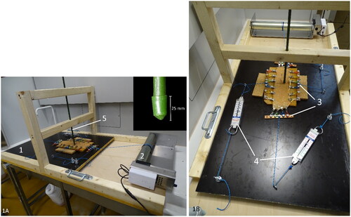
Figure 2. Histological scoring of abrasive lesions created with the ex vivo intestinal mucosal abrasion model. (a) Score 1, depression of the mucosa. Hematoxylin and eosin (HE). (b) Score 2, mucosal cleavage in the propria no dissection of the lamina muscularis mucosae (LMM) (HE). (c) Score 3, mucosal cleavage with dissection of the LMM. (d) Early-stage lesion in a cow with hemorrhagic bowel syndrome. Histologically very similar to score 3, with erosion and dissection of the LMM. Inset: this LMM splitting is even more clear with smooth muscle actin immunohistochemistry. Hemorrhage is evidently only apparent at the early-stage lesion (HE). Magnification = bar.
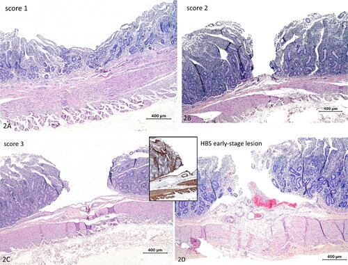
Figure 3. Hemorrhagic Bowel syndrome early-stage lesions, small intestine, bovine. (a) Two clustered erosions with minimal hemorrhage. (b) Laceration-like lesion with a detached mucosal flap and hemorrhage.
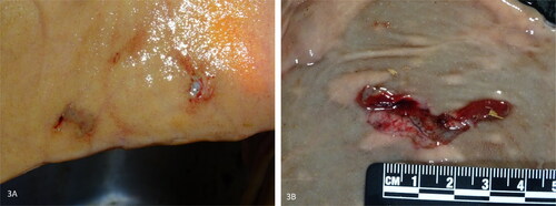
Figure 4. Diagram of gross distribution at the small intestine of early-stage lesions in hemorrhagic bowel syndrome cases (n = 7). Red and grey dots resemble early-stage lesions with and without gross hemorrhage, respectively. Lesions tend to occur more commonly proximal to the hematoma. Right to left = proximal to distal. H = hematoma.
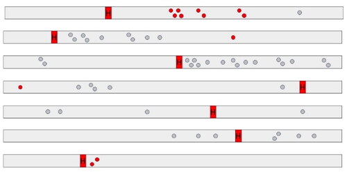
Figure 5. Lamina muscularis mucosa (LMM) at different early-stage lesions (a-c) and hematoma (d) in hemorrhagic bowel syndrome (HBS) cases. (a-b) Focally (indicated with arrows) at the ulcerated surface (asterisk) (a) to diffuse (b), degeneration of smooth muscle cells are present at the LMM. There is severe fragmentation and swelling of organelles, rarefaction and chromatin condensation. (c) De splitting of the LMM occurs through ruptured smooth muscle cells. The cell membrane of the splitted cell is indicated with arrow heads. The dissection (asterisk) cuts through the cytoplasm, adjacent to the nucleus. Inset: lower magnification overview of the splitting area of the LMM, with the area shown in ‘5C’ indicated with a rectangle. The LMM is indicated with an accolade (d) Hematoma at the start of the dissection. The cleavage of the LMM is confined to the upper portion of the inner layer of smoot muscle. Extravasated red blood cells are in between the splitted layers. TEM. Magnification = bar.
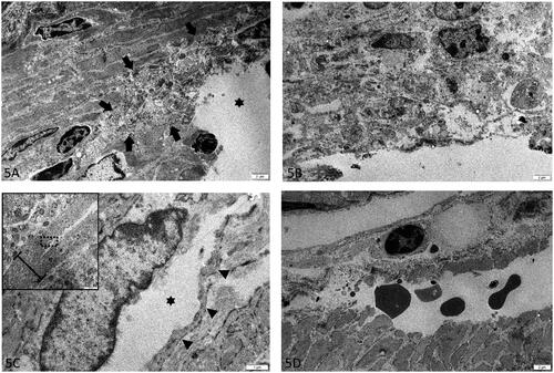
Table 1. Overview of bacterial culture of 15 early-stage lesions, in 6 cows with hemorrhagic bowel syndrome (HBS).
Figure 6. Mounted intestinal segment at the end of the experiment. Twelve injurie sites are evident. The intestine is tensed up by the gripping mechanism. The needle head color refers to the tension used for the following 4 lesions (green, 4 N; orange, 6 N; red, 8 N).

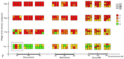
Figure 7. Summary of histological lesions scores of intestinal segments after testing with the ex vivo intestinal mucosal abrasion model. Results of 7 control animals (4 dairy and 3 beef cattle), and 3 cases of hemorrhagic bowel syndrome (HBS), are displayed. Each colored rectangle depicts results of one animal, for one combination of bi-axial tension and rod pressure. The dotted rectangle, surrounds results from 4 animals, 8 segments for the combination of 33 g and 4 N. The colors indicates the histological lesion score. Numbering of segments, as shown on the figure, are from proximal to distal.
Data availability statement
On request.