Figures & data
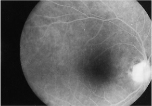
FIGURE 1 The right eye fundus exhibiting a swollen optic disc with blurred margins.
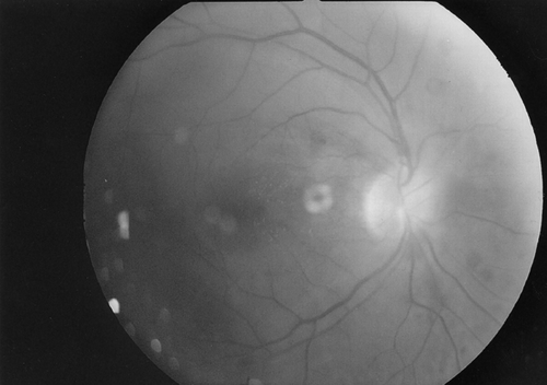
FIGURE 2 The right eye visual field showing peripheric field loss.
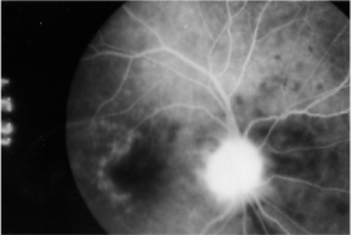
FIGURE 3 Right eye fundus fluorescein angiography revealing a hyperfluorescent optic disc on the first day.
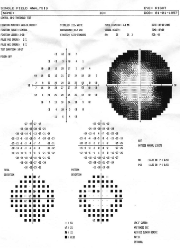
FIGURE 4 Right eye fundus fluorescein angiography after two months showing that the hyperfluorescence in the optic disc has disappeared.