Figures & data
Table 1. Patient characteristics.
Figure 1. Schematic illustration of the study protocol. A lower-lid pentagonal resection was made as part of an entropion repair procedure. A suture was placed on the medial side of the eyelid defect and connected to a dynamometer. The eyelid was then pulled laterally to create a horizontal traction force, resulting in stretching of the eyelid, using a steadily increasing force up to a maximum of 2.3 N. The procedure was filmed and eyelid perfusion was monitored with LSCI.
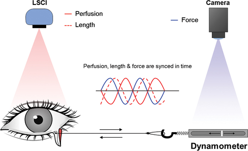
Figure 2. Representative example of intensity maps showing eyelid stretching (top) and perfusion maps (bottom) from a left lower eyelid with a pentagonal resection before and after the application of a horizontal force. Eyelid stretching (ΔL) was calculated as a percentage of the total eyelid length, from a reference point at the medial canthus. Perfusion was measured in a region of interest (dashed circle), close to the pentagonal resection, and expressed as a percent of the initial value in the relaxed eyelid. Note the decrease in perfusion (d) when the eyelid is elongated by the traction force.
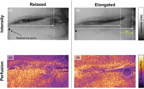
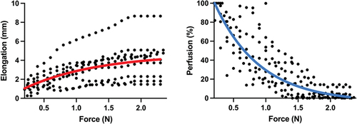
Figure 3. Eyelid stretching and blood perfusion in relation to traction force. Stretching was measured from a reference point at the medial canthus to the pentagonal resection. Blood perfusion is expressed as a percentage of the value before traction (100%). Nonlinear regression with one-phase decay was performed to describe the change in stretch and perfusion with increasing force.