Figures & data
Figure 1. Schematic diagram of the Tango system. The system comprises three components: GPCR::TCS::LexA, β-Arrestin-TEV and lexAop-reporter. Activation of GPCR by ligand binding results in phosphorylation of its C-terminus by GRK. β-Arrestin-TEV is then recruited to the phosphorylated C-terminus of GPCR::TCS::LexA and cleaves the TCS to release LexA. Released LexA translocates into the nucleus and induces reporter expression from the lexAop-reporter transgene.
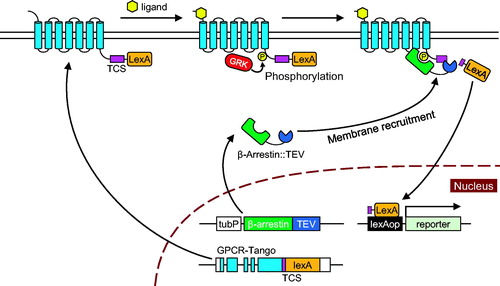
Figure 2. Targeted integration of the Tango sensor. First, a transgene cassette containing T2A-GAL4 is inserted into the C-terminus of the target GPCR by CRISPR/Cas9-mediated knock-in. This results in in-frame fusion of the GPCR and T2A-GAL4. 3xP3-was used as a visible marker for selection of transformants. It was subsequently removed by transient expression of Cre recombinase. The resultant T2A-GAL4 cassette is flanked by a phiC31 attP site and a loxP site, which are used for exchange of T2A-GAL4 for a Tango sensor by RMCE. A plasmid vector that contains the Tango sensor flanked by attB and loxP is integrated into the attP site between the GPCR and the Tango sensor. RMCE is completed by removal of the 3xP3-venus selection marker and the vector backbone by treatment with Cre recombinase.
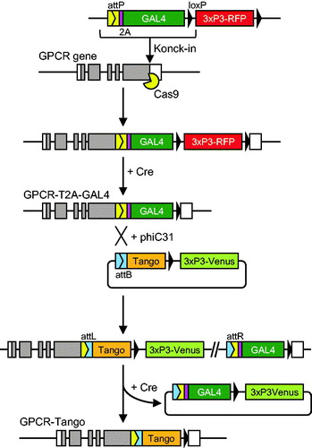
Figure 3. Spatiotemporal patterns of Tango activity in the female reproductive organ. (A) SPR-expressing cells in the female reproductive organ. SPR-expressing cells were visualized in flies that carry SPR-T2A-GAL4 and UAS-mCD8-GFP. Prominent expression was detected in the uterus (UT), the spermatheca (ST), part of the seminal receptacle (SR) and part of the oviduct (OD). No GFP expression was observed in the ovary (OV). Left: the reproductive organ of a virgin female. Right: the reproductive organ of a female fly 24 hours after mating. F-actin (magenta), mCD8-GFP (green). (B) Time-course analysis of reporter expression in SPR-Tango-L and -Tango-VL after mating. Left: Schematic designs. Each Tango sensor has a TEV cleavage site (TCS) and LexA. In addition, Tango-VL carries a V2 tail to promote β-Arrestin recruitment. Tango-L and Tango-VL carry a mutant TCS (amino-acid sequence: ENLYFQL) which is a less efficient substrate of TEV. Right: Strong mCD8-GFP was observed in the reproductive organ from 16 hours after mating in both Tango-L and Tango-VL. (C) Reporter expression in SPR-Tango-G and Tango-VG after mating. Left: Schematic designs. Tango-G and Tango-VG carry a wild-type TCS (ENLYFQG) that is cleaved by TEV by maximal efficiency. Right: Strong GFP expression was observed in the uterus and the oviduct regardless of mating status in both Tango-G and Tango-VG. The seminal receptacle and the spermatheca were removed from the reproductive organs before imaging. Scale bars in A, B, and C: 200 µm.
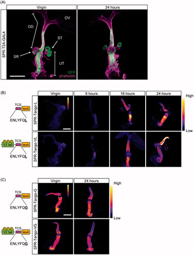
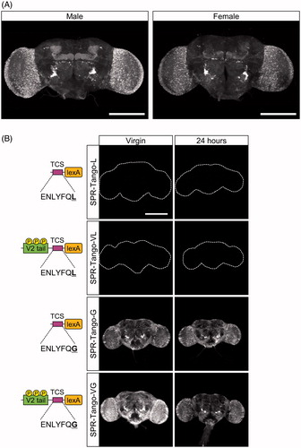
Figure 4. Spatiotemporal patterns of Tango activity in the brain. (A) SPR-expressing cells in male and female brains. SPR-expressing cells were visualized in flies that carry SPR-T2A-GAL4 and UAS-CD8-GFP. GFP was broadly expressed throughout the whole brains of both sexes. (B) Absence of mating-dependent changes in SPR-Tango activity. Left column: brains of virgin females. Right column: brains of females 24 hours after mating. The Tango variant used is indicated on the left. No GFP expression was observed in Tango-L or Tango-VL. Strong GFP signals were detected in Tango-G and Tango-VG in virgin females. No significant changes in the pattern or intensity were observed 24 hours after mating. Scale bars in A and B: 200 µm.