Figures & data
Figure 1. Schematic of a C. elegans sense organ in thehead. A typical C. elegans sense organ contains one or more sensory neurons (red) and two glial cells, called the sheath (orange) and socket (green). Most sensory neurons extend a ciliated dendritic ending through a tube-shaped pore formed by the sheath and socket glia. The socket glia secretes cuticle (gray) that forms an open pore through which chemosensory dendrite endings protrude, as shown, or a closed sheet into which mechanosensory dendrite endings are embedded (not shown). Tight junctions (yellow) are present between the neuron and sheath glia, the sheath and socket glia, and the socket glia and skin. See online version for color figure.
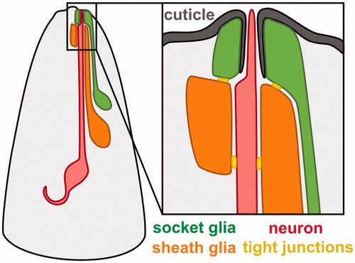
Table 1. Glial promoters used in the literature.
Figure 2. Cell-type-specific promoters for sheath and socket glia. (A) Merged brightfield images and pseudo-colored fluorescence projections of animals expressing fluorescent proteins under control of promoters selected for brightness, specificity, and consistency. F16F9.3 (pink, AMsh and PHsh); F11C7.2 (purple, AMsh only); hlh-17 (orange, CEPsh); grl-2 (blue, AMso, PHso1, and PHso2; also expressed in excretory duct and pore cells, white arrow); grl-18 (green, ILso). (B) Magnified images of AMsh (pink, F16F9.3), CEPsh (orange, hlh-17), and AMso (blue, grl-2), showing that their brightness is sufficient to resolve fine structural details including the tube-like pores of the AMsh and AMso glia and the branch-like posterior processes of the CEPsh glia. (C) Head of an animal expressing grl-18pro:GFP (green, ILso glia) and klp-6pro:mCherry (red, IL2 neurons), demonstrating that the grl-18 promoter labels the six ILso glia, identified by their processes which each form a pore for the ciliated dendritic ending of an IL2 sensory neuron. See online version for color figure.
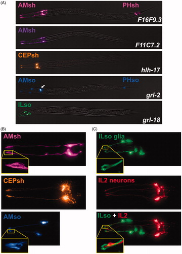
Table 2. Recommended cell-type-specific glial promoters.
Figure 3. Promoters expressed in combinations of glia. (A) Images of head expression of GFP under control of the pan-glial promoter mir-228 and the variable glial promoters delm-1 and itx-1. (B) Quantification of the number of GFP-expressing glia observed with delm-1 (n = 20) and itx-1 (n = 24) extrachromosomal reporters. Error bars indicate standard deviation. p < 0.0001, unpaired t-test with Welch’s correction. (C) Merged brightfield and fluorescence projections of a whole animal expressing pan-glial mir-228pro:GFP.
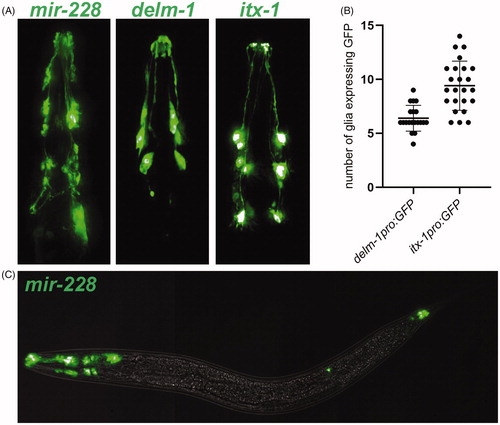
Figure 4. Plasticity in glial gene expression. (A) vap-1pro:RFP expression in AMsh glia in animals at the third larval (L3), fourth larval (L4), and 1-day adult life stages, showing developmental regulation of gene expression. (B) ver-1pro:GFP expression in AMsh glia in L4 animals grown at 15 °C, 20 °C, or 25 °C, showing thermoregulation of gene expression as previously described (Procko et al., Citation2012, Citation2011). (C) Quantification of vap-1pro:RFP fluorescence in AMsh glia from second larval (L2) stage to 1-day adults and in dyf-7(ns119) L4 and adult animals, showing that sensory defects alter gene expression. n = 15–16 per group. p < 0.05, dyf-7 L4 vs dyf-7 adults; p < 0.0001, dyf-7 adults vs wild-type adults; unpaired t-test with Welch’s correction (D) Quantification of ver-1pro:GFP fluorescence in the AMsh glia in L4 animals from 15 °C to 25 °C. n = 21–23 per group. p < 0.05, 20 °C vs 15 °C; p < 0.0001, 20 °C vs 25 °C; Brown-Forsyth and Welch one-way ANOVA with Dunnett’s multiple comparisons test.
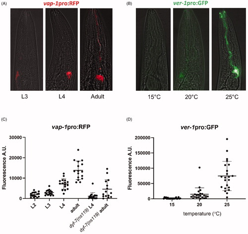
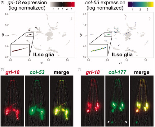
Figure 5. Prospective identification of novel cell-type-specific promoters for ILso glia. (A) Cell cluster plots illustrating the expression of grl-18 and col-53 in embryonic glia and excretory cells from Packer et al., Citation2019. Each point represents an individual cell. The color indicates the relative expression level of each gene. For grl-18, low expression is black and high expression is red. For col-53, low is blue and high is green/yellow. Gray indicates no detected transcripts for the gene of interest. Inset box, the cell cluster predicted to include ILso glia. Cell cluster plot for col-177 is in Supplementary Figure S1(A). (B, C) Merged brightfield and fluorescence images of an animal expressing grl-18pro:mApple together with (B) col-53pro:GFP or (C) col-177pro:GFP, showing that these markers are expressed in the same cells. col-177 is also faintly expressed in cells with a glial morphology, tentatively identified as OLL socket glia in the head (arrowheads, see Supplementary Figure S1(B)) and ADE and PDE socket glia in the body. See online version for color figure.