
Figures & data
Figure 1. Exosomes from macrophage promotes apoptosis of A549. A. Apoptosis assay by CaspACE FITC-VAD-FMK In Situ Marker showed CM from macrophage could promote apoptosis of A549, compared with A549 control. CM: conditioned medium from macrophage. *** P < 0.001. B. Western blot shows BCL2 downregulation in A549 cells after co-culture with CM of macrophage compared with control. CM: conditioned medium from macrophage. C. Western blot for exosomes marker CD63, actin from the lysate of cells from which exosomes were isolated as the loading control. Exo: exosomes. GW4689 is an exosome generation inhibitor. The exosomes generated by macrophage was effectively inhibited by GW4689. D. Apoptosis assay by CaspACE FITC-VAD-FMK In Situ Marker showed that exosomes isolated from macrophage could promote apoptosis of A549, compared with exosomes from macrophage treated with PBS and A549 control. *** P < 0.001. E. Western blot for BCL2 in A549, using actin as the loading control. Exo: exosomes. Each experiment was repeated at least 3 times. GW4689 is an exosome generation inhibitor. In GW4689 treated macrophage group, the BCL2 was upregulated which indicated that apoptosis was inhibited.
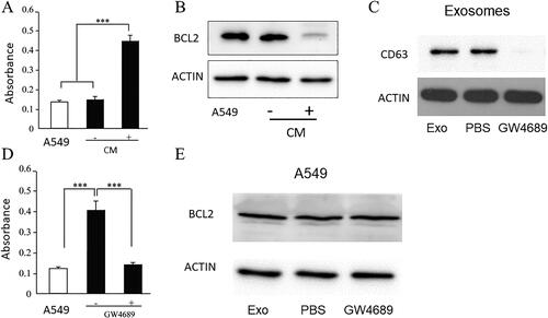
Figure 2. MiR-223 Is highly presented in exosomes and is effectively transferred into A549.
A. QPCR showed miR-223 level in macrophage, isolated exosomes and A549. *** P < 0.001. B. QPCR showed the expression of miR-223 in A549. Ctrl bar is the miR-223 level in A549 without treatment, the graph showed the expression of miR-223 in A549 increased via a dose-dependent manner. C. QPCR showed the expression of miR-223 in A54. Ctrl bar is the miR-223 level in A549 which treated with different dosages of exosomes from macrophages. Antagomir-223 bar is miR-223 level in A549 which treated with exosomes from macrophages of being transfected with Antagomir-223. Mφ: macrophage, Exo: exosomes. *** P < 0.001 each experiment was repeated at least 3 times.
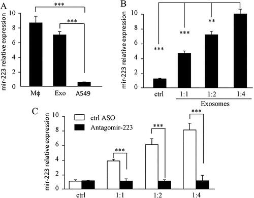
Figure 3. MiR-223 promotes endothelial cell apoptosis through targeting IGF-1R.
A. Isolated exosomes from CM of macrophage treated with antisense oligonucleotide against miR-223 couldn’t promote apoptosis of A549, CM without ASO as control. Overexpression of miR-223 in A549 promotes apoptosis of A549, empty vector as control. *** P < 0.001 EV: empty vector. ASO: antisense oligonucleotide. B. Isolated exosomes from CM of macrophage treated with antisense oligonucleotide against miR-223 couldn’t promote apoptosis of A549, CM without ASO as control. Overexpression of miR-223 in A549 promotes apoptosis of A549 as early as 12 h after incubation, empty vector as control. *** P < 0.001 EV: empty vector. ASO: antisense oligonucleotide. C. Bioinformatics analysis showed that miR-223 could bind to IGF-1R in 3’UTR. D. Western blot for IGF-1R in A549, using actin as the loading control. Exo: exosomes. EV: empty vector. E. Luciferase activity in A549 cells with co-transfection of miR-223 and empty vector or WT or mutant UTR 3’UTR of IGF-1R. *** P < 0.001. EV: empty vector. F. Apoptosis assay by CaspACE FITC-VAD-FMK In Situ Marker. A549 Cells were transfected with miR-223. Using the transfected Cells, the caspase activity of overexpression IGF1R and knock down of IGF1R were tested. The absorbance value stands for the activity of caspase and higher absorbance stands for higher activity of caspase. *** P < 0.001. SCR: Control group.
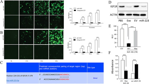
Figure 4. MiR-223 inhibits IGFR-1R/AKT/mTOR signaling pathway.
A. Western blot for AKT/mTOR signaling pathway in A549 overexpressing miR-223, using actin as the loading control. B. Western blot for AKT/mTOR signaling pathway in A549 overexpressing IGF-1R, using actin as the loading control. EV: empty vector. Each experiment was repeated at least 3 times. C. A549 apoptosis rate was detected by flow cytometry. Non-exosomes treated A549 cells used as control group. The miR-223 picture stands for the apoptosis rate of miR-223 overexpressed A549 cells and IGF-1R picture stands for the apoptosis rate of IFG-1R and miR-223 overexpressed A549 cells. The apoptosis rate was calculated by Q2 + Q3.
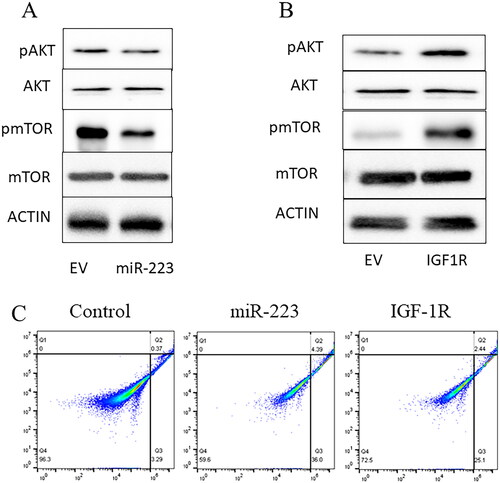
Figure 5. MiR-223 inhibits IGF-1R expression in acute lung injury.
A. Relative expression of miR-223 in the lungs of wild-type mice in a model of LPS-induced acute lung injury, treated with control anticoagulants or anticoagulants targeting miR-223 (n = 20). B. Expression of IGF1R in the lungs of wild-type mice treated with control anticoagulant or anticoagulant against miR-223 in a model of LPS-induced acute lung injury (n = 20). C. Spearman’s correlation between miR-223 and IGF1R in mouse lungs in a model of LPS-induced acute lung injury. D. Determination of eosinophils in the alveolar epithelial cell layer by light microscopic analysis of H&E-stained mouse lung sections.
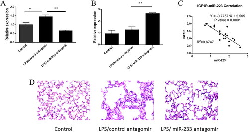
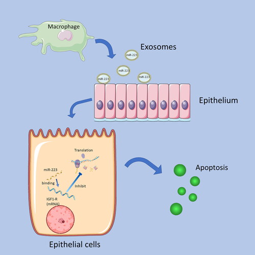
Figure 6. Summary of the mechanism of MiR-223 inhibits IGF1-R.
Exosomes of macrophages contain miR-223, and when miR-223 is transported to epithelial cells by Exosomes, the miR-223 binding to the mRNA of IGF1-R receptors and inhibits its translation. Decreased IGF1-R expression promotes epithelial cell apoptosis.
Data availability statement
The data are deposited in Figure share with DOI:10.6084/m9.figshare.23546049 and 10.6084/m9.figshare.23546199.