Figures & data
TABLE 1 Immunohistochemistry Results
Figure 1 (a) MRI showing the 13 × 10 × 6-cm biceps muscle mass. (b) MRI of the 7 × 6 × 3-cm trapezius muscle mass. Rotation of 90 °.
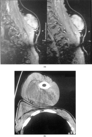
Figure 2 (a) Macrosection of the biceps muscle mass. (b) Macrosection of the trapezius muscle mass.
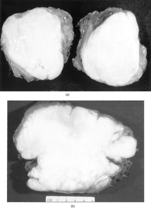
Figure 3 Sharp margins of the lesion (a) and deeply collagenized stroma with spindle to stellate cells (b).
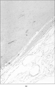
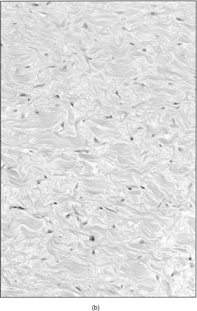
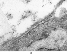
Figure 4 Fibroblasts with numerous dilated rough endoplasmic reticulum cisternae (arrows), × 3600.
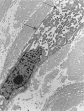
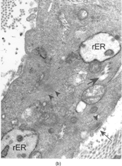
Figure 5 Portion of a myofibroblast showing intermediate filaments, actin filaments (arrowheads), dilated rough endoplasmic reticulum cisternae (rER), collagen inclusions (large and short arrows), and fibronexus (thin and long arrow), × 9,000. Insert: Detail of fibronexus junction showing a distinct fibrillary structure, × 56,000.