Figures & data
Figure 1 Microvessel lined by endothelium in the outer media wall, ×1,350.
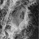
Figure 2 Small cavities (clefts) lined by normal or fragmented elastic fibers, ×550.
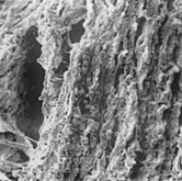
Figure 3 Pseudocyst lacking of endothelium, anfractuous, and lined by fragmented elastic fibers and smooth muscle cells with fibrous or fibrin bridges in its lumen, ×1,500.
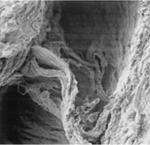
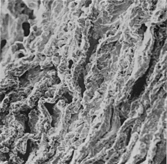
Figure 4 Elastic fibers three-dimensional disarray, fragmentation, and diastases with clefts similar to the big ones in the convexity, ×1,500.