Figures & data
Figure 1. Light micrographic findings of attached and isolated Beas-2B cells (H&E staining). (a) low-power view of attached cells. Small whirlings are noted (magnification 100х), (b) low-power view of isolated cells. Various sized cells and nuclear chromasia are noted(magnification 100х), (c) high-power view of attached cells (magnification 200х), (d) high-power view of trypsinized-isolated cells (magnification 200х), (e) large to medium sized type a cells with single and euchromatic nuclei with abundant cytoplasm, (f) medium- to small-sized type a cells with single and euchromatic or hyperchromatic nuclei, (g, h) small-type B cells with hyperchromatic nuclei with scanty dense reddish cytoplasm (arrow).
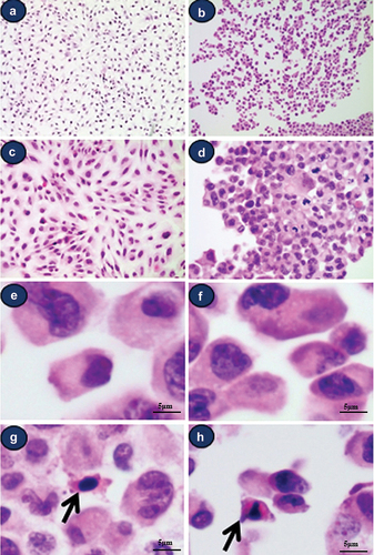
Figure 2. Light microscopic findings of various morphologically atypical cells in Beas-2B cell line. (a) atypical cell with double nuclei (b) atypical cell with multilobulated nuclei (c) atypical cell with hyperchromatic obscured nuclei, (d) multilobulated cell and mitotic figure (arrow) (a-d) (e) all cultured cells show strong positivity for Ki-67 (F) Ki-67 staining of normal human bronchial epithelial tissue; 1‒3 positive cells/100 bronchial epithelial cells.
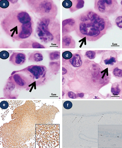
Figure 3. Ultrastructural observations of isolated cells in Beas-2B. (a) type a cell showing an indented nucleus with prominent nucleoli surrounded by a complex cytoplasm that contains prominent rough endoplasmic reticulum, Golgi apparatus, and vacuolation. (b) type B cells show a round shape with scant identifiable organelles. (c-d) abundant microvilli and intermediate filaments. (e) unspecifiable round-shaped feature (f) multilobulated atypical cells showing prominent nucleoli and abundant vacuolation.
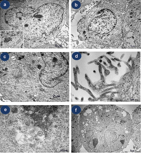
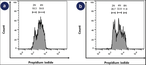
Figure 4. Result of DNA contents of Beas-2B. Flow cytometry analysis for cell cycle (a) control human IPSc cells. (b) Beas-2B cell line. Beas-2B cell line contains 59.6% the proportion of 2N and 4N, and 40.4% the proportion of abnormal karyotypes including 8N (11.9%).