Figures & data
Table 1 Clinicopathologic and Ultrastructural Findings in 15 Low-grade Myxofibrosarcomas
FIG. 1 Microscopic appearance of low-grade myxofibrosarcoma: (A) low power with spindle cells separated by abundant myxoid stroma and showing condensation around curvilinear vessels (×200;MFS6); (B) detail of a pseudo-lipoblast showing multivacuolated cytoplasm, with similar appearance as the extracellular matrix (×400;MFS15).
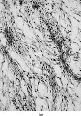
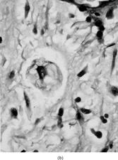
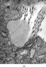
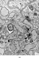
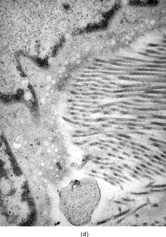
FIG. 2 Ultrastructure of myxofibrosarcoma: (A) low-power showing fusiform cells separated by myxoid stroma; abundant cytoplasm is filled with numerous dilated RER cisternae (×4, 320;MFS13);(B) 2 “pseudo-lipoblasts”-type cells showing marked distension and cystic dilatation of RER cisternae (×5, 400;MFS7);(C, D) detail of the distended cisternae, which appear either empty or filled with a granular electronuclent material (×7, 929and ×18,720; MFS 15);(E) cytoplasmic detail with abundant RER, prominent Golgi, and lysosomes (×12, 780;MFS1);(F) plump spindle cells with abundant cytoplasm rich in vimentin-type intermediate filaments, RER, and dense mitochondriae. In addition focal subectoplasmic actin condensation and short filopodia-type cell processes are also evident (×7, 920;MFS9).
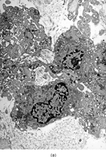
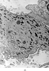
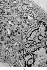
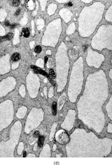
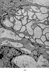

Table 2 Clinicopathologic and Ultrastructural Findings in 12 cases of Low-grade Fibromyxoid Sarcoma
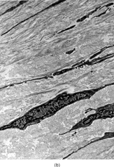
FIG. 3 Microscopic appearance of low-grade fibromyxoid sarcoma: showing bland uniform spindle cells arranged in a swirling pattern and embedded in an alternating myxoid and fibrosing stroma (×40;FMS9).
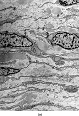
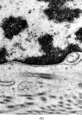
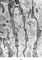
FIG. 4 Ultrastructure of fibromyxoid sarcoma: (A, B) low power showing delicate spindle cells with long, narrow and nonbranching cell processes arranged in a loose (×5, 400;FMS4) or collagenized stroma (×5, 400;FMS8);(C) detail of cell processes showing sparse organelles and small pinocytotic vesicles (×18, 720; FMS 4);(D) linear arrangement of numerous pinocytotic vesicles (×30, 420; FMS 9);(E) cytoplasmic detail with moderately developed RER as well as an ovoid fibrillary structure consistent with angulate lysosomes (×25, 200;FMS10);(F) detail of an angulate lysosomes associated with scattered dense deposits (×17, 000; FMS 12);(G) extracellular deposit of amorphous flocculent substance (×37, 800; FMS 9);(H) intracisternal collagen (×30, 000;FMS3).