Figures & data
Figure 1. The “language network”: Areas of the brain frequently activated across very different language tasks. (A) Areas activated in association with silent word generation in normal controls (Prabhakaran et al., Citation2007); (B) areas of activation associated with word retrieval (naming and oral reading minus saying, “1, 2, 3”; Parker Jones et al., Citation2012); (C) areas of activation associated with syntax repetition (Segaert et al., Citation2012); and (D) areas of activation associated with reading (blue) and spelling (green; Rapp & Lipka, Citation2011). [To view this figure in colour, please see the online version of this journal.]
![Figure 1. The “language network”: Areas of the brain frequently activated across very different language tasks. (A) Areas activated in association with silent word generation in normal controls (Prabhakaran et al., Citation2007); (B) areas of activation associated with word retrieval (naming and oral reading minus saying, “1, 2, 3”; Parker Jones et al., Citation2012); (C) areas of activation associated with syntax repetition (Segaert et al., Citation2012); and (D) areas of activation associated with reading (blue) and spelling (green; Rapp & Lipka, Citation2011). [To view this figure in colour, please see the online version of this journal.]](/cms/asset/21a68f6f-a175-4bf0-a11a-76bb10b5df5f/pcgn_a_875467_f0001_c.jpg)
Table 1. Participant 1 performance on the Johns Hopkins lexical battery
Table 2. Western Aphasia Battery and additional tasks
Figure 2. (A) Participant 1's DWI (left) and PWI (right) before intervention to restore blood flow shows hypoperfusion of left frontal and inferior temporal cortex, and acute infarcts in subcortical watershed areas. TTP = time-to-peak. (B) DWI and PWI post intervention shows reperfusion of left frontal and inferior temporal cortex (and unchanged acute subcortical infarcts). (C) Language testing shows improvement in oral picture naming and oral naming of objects from tactile input after reperfusion of left frontal and inferior temporal cortex. [To view this figure in colour, please see the online version of this journal.]
![Figure 2. (A) Participant 1's DWI (left) and PWI (right) before intervention to restore blood flow shows hypoperfusion of left frontal and inferior temporal cortex, and acute infarcts in subcortical watershed areas. TTP = time-to-peak. (B) DWI and PWI post intervention shows reperfusion of left frontal and inferior temporal cortex (and unchanged acute subcortical infarcts). (C) Language testing shows improvement in oral picture naming and oral naming of objects from tactile input after reperfusion of left frontal and inferior temporal cortex. [To view this figure in colour, please see the online version of this journal.]](/cms/asset/ef8bfb3b-2065-45f6-ab80-68e24d0d7e6f/pcgn_a_875467_f0002_c.jpg)
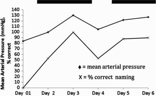
Figure 3. Temporal correlation between mean arterial pressure and oral picture naming accuracy for Participant 1. The two solid lines at the top of represent times when Participant 1 was receiving intervention to increase blood pressure.

Figure 4. DWI (Panel A) and apparent diffusion coefficient (ADC; Panel B) sequences show a subcentimetre area of diffusion restriction in the left thalamus for Participant 2. Functional magnetic resonance imaging (fMRI) data registered in Montreal Neurological Institute (MNI) space shows areas of activation associated with silent word generation at Day 3 (Panel C) and 8 weeks (Panel D). fMRI activation maps are false discovery rate (FDR) corrected for multiple comparisons and are displayed with a threshold of p < .05. A difference statistical map (Panel E) shows activation that is greater at 8 weeks than 3 weeks in yellow, and activation that is greater at 3 weeks than 8 weeks in blue. This last map shows change in statistical maps as a function of time. [To view this figure in colour, please see the online version of this journal.]
![Figure 4. DWI (Panel A) and apparent diffusion coefficient (ADC; Panel B) sequences show a subcentimetre area of diffusion restriction in the left thalamus for Participant 2. Functional magnetic resonance imaging (fMRI) data registered in Montreal Neurological Institute (MNI) space shows areas of activation associated with silent word generation at Day 3 (Panel C) and 8 weeks (Panel D). fMRI activation maps are false discovery rate (FDR) corrected for multiple comparisons and are displayed with a threshold of p < .05. A difference statistical map (Panel E) shows activation that is greater at 8 weeks than 3 weeks in yellow, and activation that is greater at 3 weeks than 8 weeks in blue. This last map shows change in statistical maps as a function of time. [To view this figure in colour, please see the online version of this journal.]](/cms/asset/cd5f9c1b-b984-4407-8e4d-afcc617208e0/pcgn_a_875467_f0004_c.jpg)
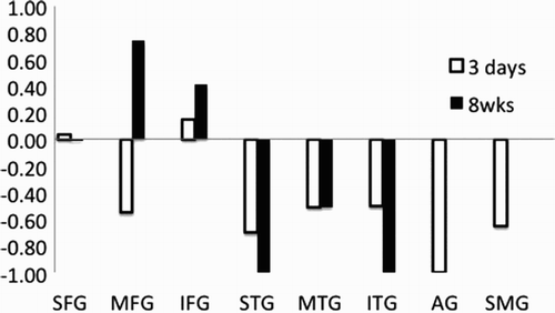
Figure 5. Lateralization index for word generation in eight regions of interest (ROIs) at 3 days compared to 8 weeks in Participant 2. ROIs are: superior frontal gyrus (SFG), medial frontal gyrus (MFG), inferior frontal gyrus (IFG), superior temporal gyrus (STG), middle temporal gyrus (MTG), inferior temporal gyrus (ITG), supramarginal gyrus (SMG), and angular gyrus (AG).

Figure 6. Panel A: DWI and PWI at Day 1 for Participant 3. Panel B: Activation associated with picture naming for Participant 3 at Day 3. Panel C: Activation associated with picture naming at 8 weeks. Functional magnetic resonance imaging (fMRI) activation maps are false discovery rate (FDR) corrected for multiple comparisons and are displayed with a threshold of p < .05. Panel D: A difference statistical map shows activation that is greater at 8 weeks than 3 weeks in yellow, and activation that is greater at 3 weeks than 8 weeks in blue. [To view this figure in colour, please see the online version of this journal.]
![Figure 6. Panel A: DWI and PWI at Day 1 for Participant 3. Panel B: Activation associated with picture naming for Participant 3 at Day 3. Panel C: Activation associated with picture naming at 8 weeks. Functional magnetic resonance imaging (fMRI) activation maps are false discovery rate (FDR) corrected for multiple comparisons and are displayed with a threshold of p < .05. Panel D: A difference statistical map shows activation that is greater at 8 weeks than 3 weeks in yellow, and activation that is greater at 3 weeks than 8 weeks in blue. [To view this figure in colour, please see the online version of this journal.]](/cms/asset/6871d33f-7d1e-438e-af29-e06003f1af44/pcgn_a_875467_f0006_c.jpg)
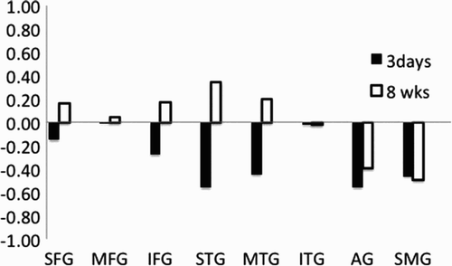
Figure 7. Lateralization index for picture naming in eight regions of interest (ROIs) at 3 days compared to 8 weeks in Participant 3. ROIs are: superior frontal gyrus (SFG), medial frontal gyrus (MFG), inferior frontal gyrus (IFG), superior temporal gyrus (STG), middle temporal gyrus (MTG), inferior temporal gyrus (ITG), supramarginal gyrus (SMG), and angular gyrus (AG).

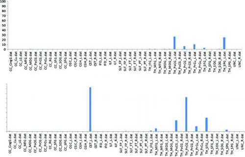
Figure 8. Probabilistic fibre tract lesion disruption analysis. The bar graph illustrates the percentage of disruption to each white matter tract for Participant 2 (top panel) and Participant 3 (lower panel). CST_L, cortico spinal cord (left); PrCG_L = precentral gyrus (left).

Figure 9. Global analysis of cortical function disruption in Participant 3. The degree of white matter tract disruption in each cortical region is determined from the intersection of the lesion region of interest (ROI) and a probabilistic fibre tract atlas. The results are presented as a percentage of total white matter tract disruption, normalized to the region of highest level of disruption, in Participant 3. [To view this figure in colour, please see the online version of this journal.]
![Figure 9. Global analysis of cortical function disruption in Participant 3. The degree of white matter tract disruption in each cortical region is determined from the intersection of the lesion region of interest (ROI) and a probabilistic fibre tract atlas. The results are presented as a percentage of total white matter tract disruption, normalized to the region of highest level of disruption, in Participant 3. [To view this figure in colour, please see the online version of this journal.]](/cms/asset/33e1119b-a01e-44e2-a815-d03288576087/pcgn_a_875467_f0009_c.jpg)
Figure 10. Panel A: DWI (top) and PWI (bottom) at Day 1 for Participant 4. Panel B: DWI (top) and PWI (bottom) at Day 1 for Participant 5. Panel C: Functional magnetic resonance imaging (fMRI) activation associated with the orthographic task (retrieval of spelling) for Participant 4. Panel D: fMRI activation associated with the orthographic task (retrieval of spelling) for Participant 5. Panel E: Activation for Participant 4 minus activation for Participant 5, associated with the orthographic task. fMRI activation maps are false discovery rate (FDR) corrected for multiple comparisons and are displayed with a threshold of p < .05. Volumes with greater than 2 mm movement were censored out. [To view this figure in colour, please see the online version of this journal.]
![Figure 10. Panel A: DWI (top) and PWI (bottom) at Day 1 for Participant 4. Panel B: DWI (top) and PWI (bottom) at Day 1 for Participant 5. Panel C: Functional magnetic resonance imaging (fMRI) activation associated with the orthographic task (retrieval of spelling) for Participant 4. Panel D: fMRI activation associated with the orthographic task (retrieval of spelling) for Participant 5. Panel E: Activation for Participant 4 minus activation for Participant 5, associated with the orthographic task. fMRI activation maps are false discovery rate (FDR) corrected for multiple comparisons and are displayed with a threshold of p < .05. Volumes with greater than 2 mm movement were censored out. [To view this figure in colour, please see the online version of this journal.]](/cms/asset/a821d3d7-0c6e-47e6-a7f8-29dbfae0fffc/pcgn_a_875467_f0010_c.jpg)
Table 3. Number of voxels with activation in nine regions of interest in each hemisphere
Figure 11. Activation associated with reading for Participant 5. Functional magnetic resonance imaging (fMRI) data are adjusted for multiple corrections and are presented with threshold of p < .05. [To view this figure in colour, please see the online version of this journal.]
![Figure 11. Activation associated with reading for Participant 5. Functional magnetic resonance imaging (fMRI) data are adjusted for multiple corrections and are presented with threshold of p < .05. [To view this figure in colour, please see the online version of this journal.]](/cms/asset/21c8ae39-d3cd-49c6-9bc9-8118bca4da23/pcgn_a_875467_f0011_c.jpg)