299
Views
1
CrossRef citations to date
0
Altmetric
Original

Heat-inducible amplifier vector for high-level expression of granulocyte-macrophage colony-stimulating factor

, , , , &
Pages 407-419 | Received 01 Nov 2005, Accepted 11 Apr 2006, Published online: 09 Jul 2009

Figures & data

Figure 1. Map of the GM-CSF expression constructs. (a) The first transcriptional unit consists of the inducible HSP70B promoter that drives the expression of the first gene, the transcription-activating factor tat. The second transcriptional unit contains the gene encoding GM-CSF driven by the HIV2 promoter (HIV2 LTR). (b) The HSP70B promoter directly controls the GM-CSF gene expression. (c) The constitutive CMV promoter directly drives the expression of the GM-CSF gene.
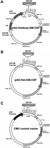
Figure 1. Map of the GM-CSF expression constructs. (a) The first transcriptional unit consists of the inducible HSP70B promoter that drives the expression of the first gene, the transcription-activating factor tat. The second transcriptional unit contains the gene encoding GM-CSF driven by the HIV2 promoter (HIV2 LTR). (b) The HSP70B promoter directly controls the GM-CSF gene expression. (c) The constitutive CMV promoter directly drives the expression of the GM-CSF gene.

Figure 2. Survival of B16 cells after heat shock. Cells were heat-shocked in a water bath at indicated times and temperatures 24 h after seeding. After another 24 h, cells were harvested and viable cells counted. Heating for 30 min at 42°C showed no significant difference from 37°C control in cell viability (p = 0.24). All conditions were repeated in triplicates with mean values shown (±SEM).
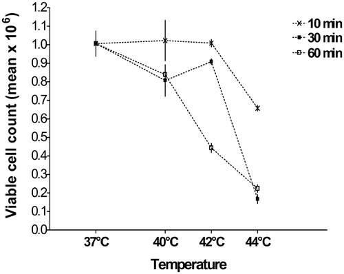
Figure 2. Survival of B16 cells after heat shock. Cells were heat-shocked in a water bath at indicated times and temperatures 24 h after seeding. After another 24 h, cells were harvested and viable cells counted. Heating for 30 min at 42°C showed no significant difference from 37°C control in cell viability (p = 0.24). All conditions were repeated in triplicates with mean values shown (±SEM).

Figure 3. Inducible gene expression using HSP70B promoter. Plasmids using the HSP70B promoter (a) showed low background activity at 37°C but large increases in luciferase expression upon heat shock compared to the CMV promoter (b). The maximum increase in HSP70B promoter-controlled luciferase expression of 250-fold was observed with heat shock of 44°C for 30 min. A heat shock of 42°C for 30 min provided an increase in luciferase activity by more than 60-fold over background. Values shown are means of relative light units (RLU) (±SEM) of triplicate experiments.
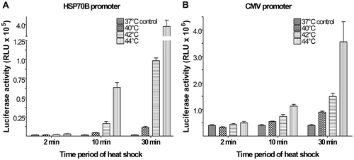
Figure 3. Inducible gene expression using HSP70B promoter. Plasmids using the HSP70B promoter (a) showed low background activity at 37°C but large increases in luciferase expression upon heat shock compared to the CMV promoter (b). The maximum increase in HSP70B promoter-controlled luciferase expression of 250-fold was observed with heat shock of 44°C for 30 min. A heat shock of 42°C for 30 min provided an increase in luciferase activity by more than 60-fold over background. Values shown are means of relative light units (RLU) (±SEM) of triplicate experiments.

Figure 4. GM-CSF production levels. Cells were heat-shocked at 42°C for 30 min 24 h post-transfection (▪) or kept at 37°C as controls (□). Supernatants were harvested 24, 48 and 72 h after transfection. GM-CSF levels in the supernatants were assayed using ELISA. Each bar represents the GM-CSF production within a 24 h period. Values shown are means of triplicate experiments (±SEM). The GM-CSF levels, 24 h after heat shock, produced by pAD-HotAmp-GM-CSF were compared to pAD-Hot-GM-CSF (p < 0.001) and the CMV control vector (p < 0.001) and to the non-heat-shocked control of pAD-HotAmp-GM-CSF (p < 0.001).
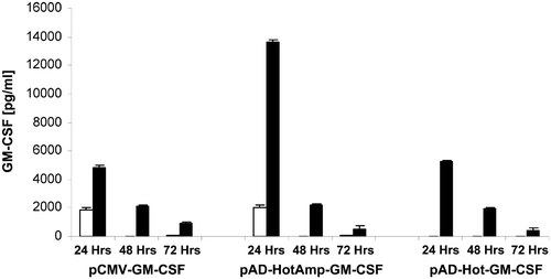
Figure 4. GM-CSF production levels. Cells were heat-shocked at 42°C for 30 min 24 h post-transfection (▪) or kept at 37°C as controls (□). Supernatants were harvested 24, 48 and 72 h after transfection. GM-CSF levels in the supernatants were assayed using ELISA. Each bar represents the GM-CSF production within a 24 h period. Values shown are means of triplicate experiments (±SEM). The GM-CSF levels, 24 h after heat shock, produced by pAD-HotAmp-GM-CSF were compared to pAD-Hot-GM-CSF (p < 0.001) and the CMV control vector (p < 0.001) and to the non-heat-shocked control of pAD-HotAmp-GM-CSF (p < 0.001).

Reprints and Corporate Permissions

Please note: Selecting permissions does not provide access to the full text of the article, please see our help page How do I view content?

To request a reprint or corporate permissions for this article, please click on the relevant link below:

Academic Permissions

Please note: Selecting permissions does not provide access to the full text of the article, please see our help page How do I view content?

Obtain permissions instantly via Rightslink by clicking on the button below:

If you are unable to obtain permissions via Rightslink, please complete and submit this Permissions form. For more information, please visit our Permissions help page.