Figures & data
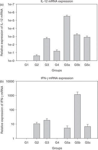
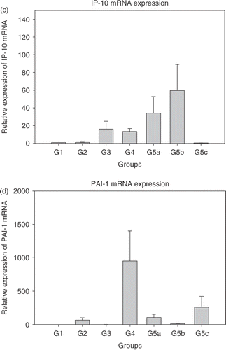
Figure 1. Relative expression of mRNAs in the treated groups 2–5c normalized to group 1 (each bar represents samples from five mice each in triplicate ± SEM). (a) Interleukin-12 (IL-12); (b) Interferon-γ (IFN-γ); (c) Interferon inducible protein-10 (IP-10); (d) Plasminogen activator inhibitor (PAI-1); and (e) Vascular endothelial growth factor (VEGF). Group 1: Normal saline injection, Control. Tumour and tissue samples collected at the same time as treatment group (5 days post-HT); Group 2: Ad LacZ + HT. Tumour and tissue samples collected 5 days post-HT; Group 3: AdhspmIL-12, no HT. Tumour and tissue samples collected at the same time as treatment group (5 days post- HT); Group 4: Normal saline injection, HT only. Tumour and tissue samples collected 5 days post-HT; Group 5: AdhspmIL-12 + HT (Group 5a: tumour sampled 6 h post-HT; Group 5b: tumour sampled 24 h post-HT; Group 5c: tumour sampled 5 days post-HT).
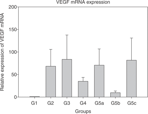
Figure 2. Tumour growth curves. The relative tumour volumes for the treatment groups are shown. A statistically significant difference was noted between the control and virus + hyperthermia groups (p = 0.017) and between AdhspmIL-12 alone vs. AdhspmIL-12 + HT groups (p = 0.02). However, on pairwise comparisons using the Kruskal-Wallis test no significant difference was found between any other group.
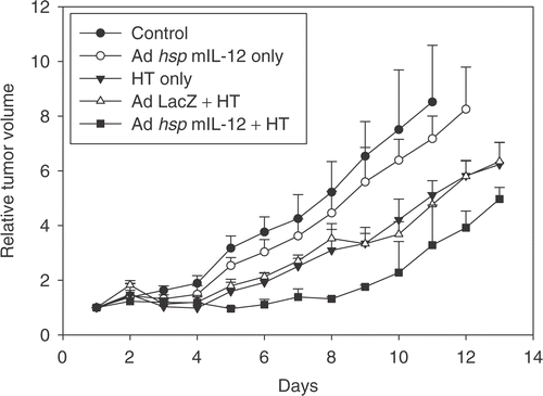
Figure 3. Tomato lectin-FITC and CD31 & hypoxyprobe staining. (a) Tomato lectin-FITC. (b) Same coordinates on the slide imaged for CD31 and hypoxyprobe staining. Area stained by CD31 is larger than FITC indicative of greater presence of the antigen than is actually patent functional vasculature. (c) Image from a control tumour section showing the patent vasculature in green. (d) IL-12 treated tumour 5 days post-hyperthermia showing reduced vasculature. (e) Section from a control group tumour showing large areas staining positive for CD31 antigen with small areas of hypoxia. (f) Section from a treated group tumour showing reduced CD31 staining and larger areas of hypoxia.
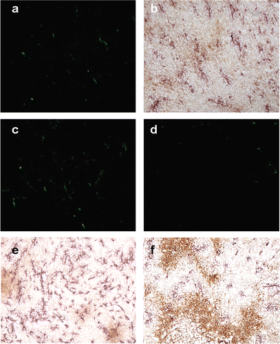
Figure 4. Area occupied by FITC and CD31 staining. (a) Percentage of field positive for Tomato lectin-FITC fluorescence, suggestive of functional vasculature. (b) Percentage of field staining positive for CD31 vasculature. The groups mentioned in the graphs are as follows: (1) Normal saline injection, Control. Tumours collected at the same time as treatment group (5 days post-HT); (2) Ad LacZ + HT. 5 days post-HT; (3) AdhspmIL-12, no HT. Same time as treatment group (5 days post-HT); (4) Normal saline injection, HT only. 5 days post-HT; (5) AdhspmIL-12 + HT. 5 days post-HT. Reduction in tumour vasculature was statistically significant (p < 0.05) in group 5 as compared to all other groups and for group 3 when compared to Control group (group 1).
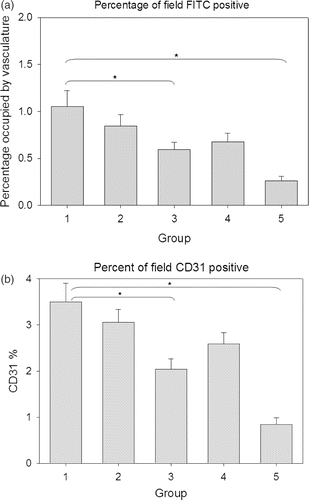
Figure 5. Area occupied by hypoxyprobe staining. The hypoxic area was higher for the AdhspmIL-12 + HT treated group as compared to all other groups. The groups mentioned in the graphs are as follows: (1) Normal saline injection, Control. Tumours collected at the same time as treatment group (5 days post- HT); (2) Ad LacZ + HT. 5 days post-HT; (3) AdhspmIL-12, no HT. Same time as treatment group (5 days post-HT); (4) Normal saline injection, HT only. 5 days post-HT; (5) AdhspmIL-12 + HT. 5 days post-HT.

Figure 6. Interleukin-12 staining. The red circles outline some of the areas staining positive for IL-12 in a haematoxylin background in the AdhspmIL-12 + HT group. This image is from one of the tumours collected 5 days post HT (100×). In the control, Ad LacZ + HT, AdhspmIL-12 alone and HT alone groups no IL-12 was detected in the immunohistochemically stained tumour sections.
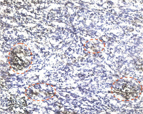
Figure 7. Lung metastases. Metastases were detected in the lungs from the mice in the control group. The metastases were seen as sheets of tumour cells replacing the normal lung architecture. (a) Lung metastasis at 40× magnification. (b) Lung metastasis at 200× magnification.
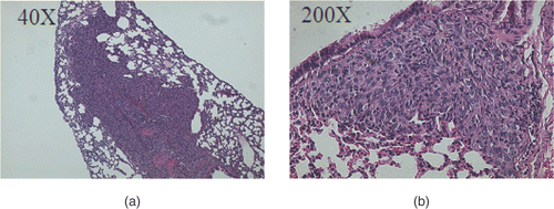
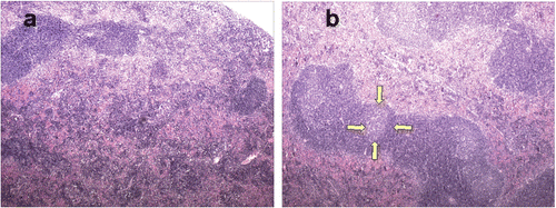
Figure 8. Spleen histology. (a) Spleen from a control group mouse (40×). (b) Spleen from a mouse euthanized 5 days post-HT showing enlarged germinal centres (the yellow arrows outline one such germinal centre). Many follicles appear to be coalescing together.